Review
. 2017 Dec;13(12):731-749.
doi: 10.1038/nrneph.2017.148. Epub 2017 Oct 30.

Isolation and characterization of urinary extracellular vesicles: implications for biomarker discovery

Affiliations
Review

Isolation and characterization of urinary extracellular vesicles: implications for biomarker discovery

Michael L Merchant et al. Nat Rev Nephrol. 2017 Dec.

Abstract

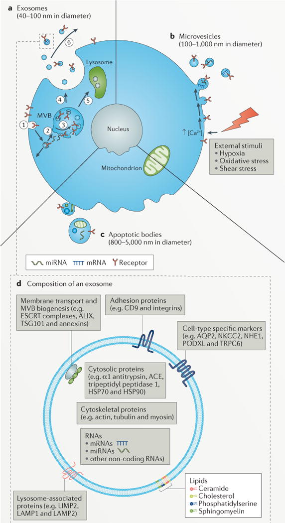
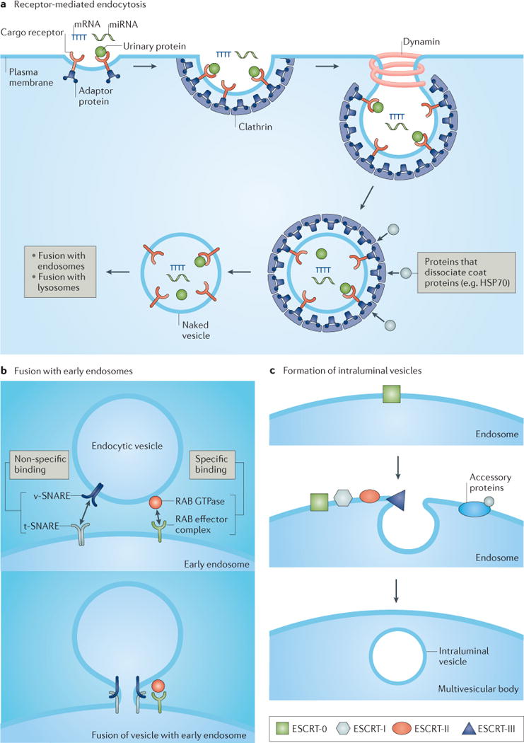
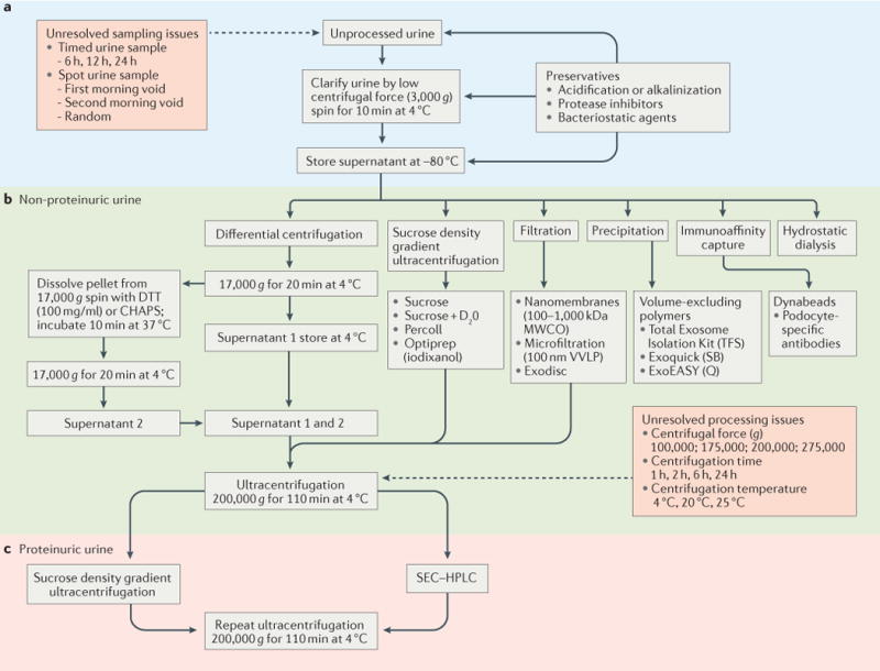
Urine is a valuable diagnostic medium and, with the discovery of urinary extracellular vesicles, is viewed as a dynamic bioactive fluid. Extracellular vesicles are lipid-enclosed structures that can be classified into three categories: exosomes, microvesicles (or ectosomes) and apoptotic bodies. This classification is based on the mechanisms by which membrane vesicles are formed: fusion of multivesicular bodies with the plasma membranes (exosomes), budding of vesicles directly from the plasma membrane (microvesicles) or those shed from dying cells (apoptotic bodies). During their formation, urinary extracellular vesicles incorporate various cell-specific components (proteins, lipids and nucleic acids) that can be transferred to target cells. The rigour needed for comparative studies has fueled the search for optimal approaches for their isolation, purification, and characterization. RNA, the newest extracellular vesicle component to be discovered, has received substantial attention as an extracellular vesicle therapeutic, and compelling evidence suggests that ex vivo manipulation of microRNA composition may have uses in the treatment of kidney disorders. The results of these studies are building the case that urinary extracellular vesicles act as mediators of renal pathophysiology. As the field of extracellular vesicle studies is burgeoning, this Review focuses on primary data obtained from studies of human urine rather than on data from studies of laboratory animals or cultured immortalized cells.


Conflict of interest statement

Competing interests statement

The authors declare no competing interests.

Figures

Figure 1
Figure 1. Mechanisms of urinary extracellular vesicle formation regulate their composition
a | Exosomes are small bilayered vesicles (40–100 nm in diameter) that contain proteins, lipids and small molecules that are derived from the plasma membrane, and/or RNA and proteins that are derived from the cytoplasm. In the first step of exosome formation, membrane proteins are internalized (endocytosed) by cells (step 1), resulting in the formation of early endosomes. As these early endosomes mature into late endosomes (step 2), invagination of their delimiting membrane produces intraluminal vesicles, by processes regulated by four distinct endosomal sorting complexes required for transport (ESCRT) complexes (step 3); these late endosomes containing intraluminal vesicles are then referred to as multivesicular bodies (MVBs). MVBs can fuse with the plasma membrane, resulting in the release of intraluminal vesicles (step 4), which are now termed exosomes. Alternatively, MVBs can fuse with lysosomes, which results in the degradation of the contents of the MVB (step 5). Exosomes can participate in molecular signalling events after their release into the urinary space or into the parenchymal interstitial space (step 6). b | Microvesicles are large bilayered vesices (100–1,000 nm in diameter) that contain plasma membrane lipids and proteins, and cytoplasmic lipids, proteins and nucleic acids. Microvesicles form when a stimulus (for example, hypoxia, oxidative stress or shear stress) drives intracellular events such as those mediated by Ca2+ or phospholipid-binding proteins, which cause the shedding and release of microvesicles from the plasma membrane. c | Apoptotic bodies are large bilayered vesicles (800–5,000 nm in diameter) that are highly heterogeneous in both size and composition. The delimiting membrane of apoptotic bodies contains plasma membrane-derived lipids and proteins and encloses cytoplasmic material that includes organelle-specific proteins (for example, those from the nucleus, mitochondria, and so on), nucleic acids and lipids. d | Composition of an exosome. Exosomes contain lipids, nucleic acids and proteins, some of which are unique to the cell type from which the exosomes form. Phospholipids and sterols, such as ceramide, sphingomyelin, phosphatidylserine and cholesterol, are important for the mechanistic and biophysical aspects of bilayer formation, curvature and fluidity, which affect membrane fusion. The mechanistic role for specific lipids (for example, phosphatidylserine) is evident from the redistribution of these lipids between inner and outer leaflets or spatial segregation into the outer leaflet of extracellular vesicles. The membrane of urinary extracellular vesicles also contain integral membrane proteins and membrane-associated proteins, such as adhesion proteins (CD9 and integrins), membrane transport and/or fusion proteins and proteins involved in MVB biogenesis (ESCRT proteins, ALG2-interacting protein X (ALIX), tumour susceptibility gene 101 protein (TSG101) and annexins), lysosomal proteins (lysosome membrane protein 2 (LIMP2), lysosome-associated membrane protein 1 (LAMP1) and LAMP2), whereas the lumen contains soluble proteins (such as α1 antitrypsin, angiotensin-converting enzyme (ACE), tripeptidyl peptidase 1 and the heat shock proteins HSP70 and HSP90), and cytoskeletal proteins (such as actin, tubulin and myosin). Nucleic acids present in the lumen of urinary extracellular vesicles include mRNAs, microRNAs (miRNAs) and long, non-coding RNAs. AQP2, aquaporin 2; NHE1, sodium/hydrogen exchanger 1; NKCC2, Na-K-2Cl cotransporter; PODXL, podocalyxin; TRPC6, short transient receptor potential channel 6.
Figure 2
Figure 2. Overview of exosome formation
Exosome formation occurs through a multistep process that is initiated by pinocytosis (not shown) or receptor-mediated endocytosis (part a), which involves the binding of urinary proteins to the apical membrane and their internalization, a process that requires the coat protein clathrin and the ATPase dynamin. Invagination of the lipid bilayer results in formation of a small unilamellar vesicle. Proteins such as heat shock protein 70 (HSP70) can dissociate coat proteins (for example, clathrin) to yield a naked vesicle that can fuse with early endosomes (part b), a process that is mediated by soluble N-ethylmaleimide-sensitive factor attachment protein receptors (SNAREs) and small RAB effector proteins. Intraluminal vesicles form after invagination of the endosomal membrane (part c), a process that is carried out by tetraspanins (not shown) and endosomal sorting complex required for transport (ESCRT) protein complexes, such as ESCRT-0 (which is responsible for cargo clustering), ESCRT-I and ESCRT-II (both are responsible for inducing bud formation), and ESCRT-III (which promotes intraluminal budding of vesicles in endosomes and vesicle scission). The dissociation and recycling of the ESCRT machinery is carried out by accessory proteins. miRNA, microRNA; t-SNARE, target SNARE; v-SNARE, vesicle SNARE.
Figure 3
Figure 3. Comparison of approaches to isolate extracellular vesicles from the urine of healthy individuals and from patients with nephrotic syndrome
Urine is collected as a spot or timed urine sample (part a). The process of urinary extracellular vesicle isolation begins with a low speed and/or low centrifugal force (3,000 g) centrifugation step for a short time (≤10 min) and at low temperature (4°C) to clarify the urine (that is, remove the flocculent material, which can include bacteria and cells). Ideally, the urine is carried forward (part b) through to the urinary extracellular vesicle isolation step. This step represents a dynamic field of investigation, and includes methods such as differential centrifugation or ultracentrifugation, single step centrifugation using density gradient material (sucrose, Percoll), filtration or ultrafiltration, precipitation (for example, Exoquick), immunoaffinity capture and hydrostatic dialysis. The complex composition of urine from patients with nephrotic syndrome interferes with the isolation of urinary extracellular vesicles, and additional steps (such as density gradient centrifugation or size exclusion chromatography (SEC)) are required (part c) to further purify the urinary extracellular vesicles from contaminating high-molecular-weight protein complexes (such as albumin) that co-isolate with the urinary extracellular vesicles. CHAPS, 3-[(3-cholamidopropyl)dimethylammonio]-1-propanesulfonate; D2O, deuterium oxide; DTT, dithiothreitol; HPLC, high performance liquid chromatography; MWCO, molecular weight cut-off; Q, Qiagen; SB, Systems Biosciences; TFS, ThermoFisher Scientific; VVLP, hydrophilic polyvinylidene difluoride.

References

    1. Wolf P. The nature and significance of platelet products in human plasma. Br J Haematol. 1967;13:269–288. - PubMed
    1. Taylor DD, Doellgast GJ. Quantitation of peroxidase-antibody binding to membrane fragments using column chromatography. Anal Biochem. 1979;98:53–59. - PubMed
    1. Ratajczak J, Wysoczynski M, Hayek F, Janowska-Wieczorek A, Ratajczak MZ. Membrane-derived microvesicles: important and underappreciated mediators of cell-to-cell communication. Leukemia. 2006;20:1487–1495. - PubMed
    1. Street JM, et al. Exosomal transmission of functional aquaporin 2 in kidney cortical collecting duct cells. J Physiol. 2011;589:6119–6127. - PMC - PubMed
    1. Valadi H, et al. Exosome-mediated transfer of mRNAs and microRNAs is a novel mechanism of genetic exchange between cells. Nat Cell Biol. 2007;9:654–659. - PubMed