Case Reports
. 2017 Jul 31:11:617-632.
doi: 10.2174/1874325001711010617. eCollection 2017.

Arthroscopic Lateral Ligament Repair Through Two Portals in Chronic Ankle Instability

Affiliations
Case Reports

Arthroscopic Lateral Ligament Repair Through Two Portals in Chronic Ankle Instability

Jorge Pablo Batista et al. Open Orthop J. .

Abstract

Objectives: Injury to the lateral ligament complex of the ankle is one of the most common sports-related injury. Usually lateral ankle evolves with excellent clinical recovery with non surgical treatment, however, near about 30% develop a lateral chronic instability sequela. Several open and arthroscopic surgical techniques have been described to treat this medical condition.

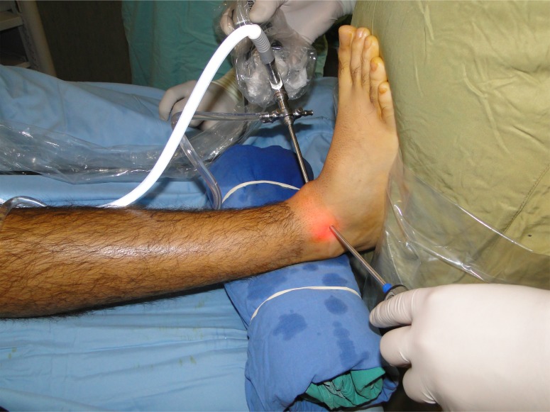
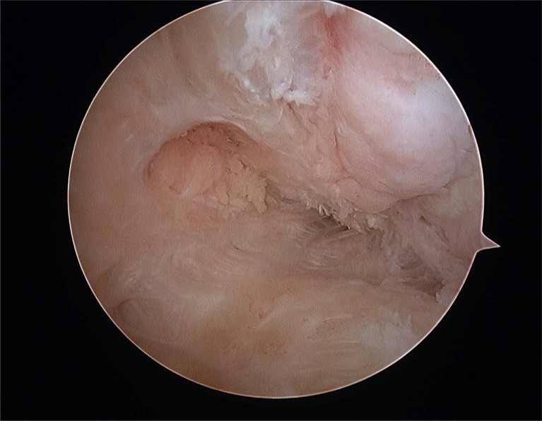
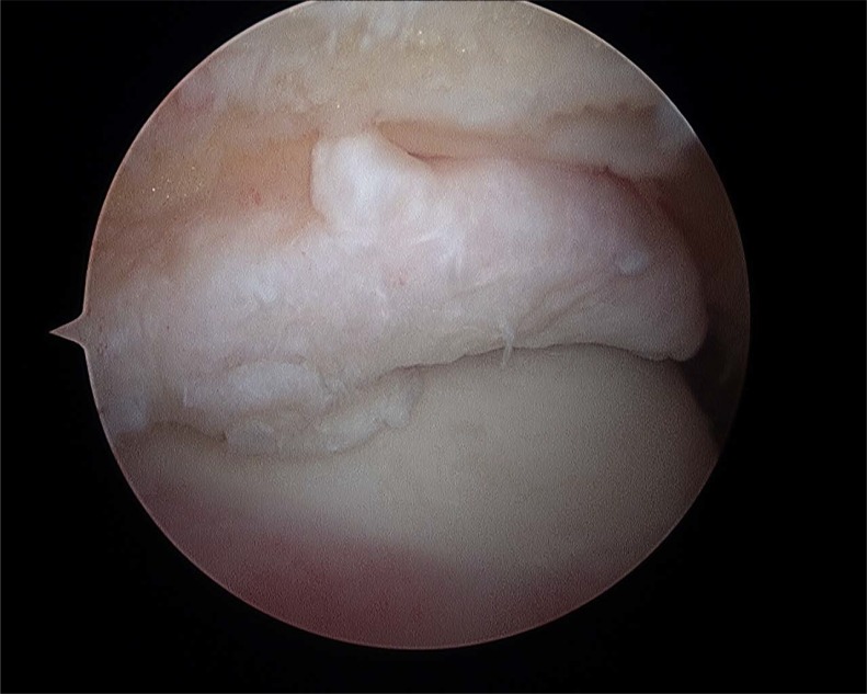
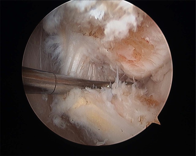
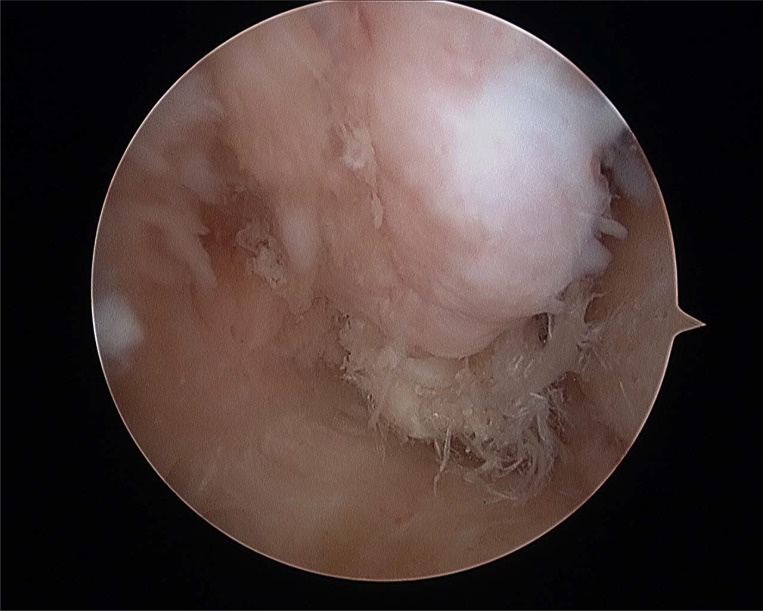
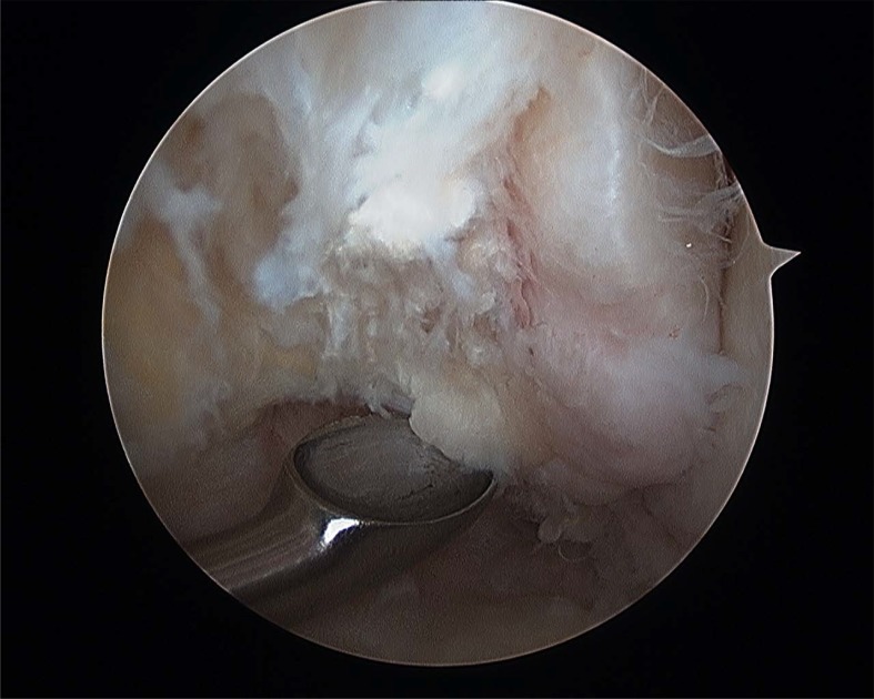
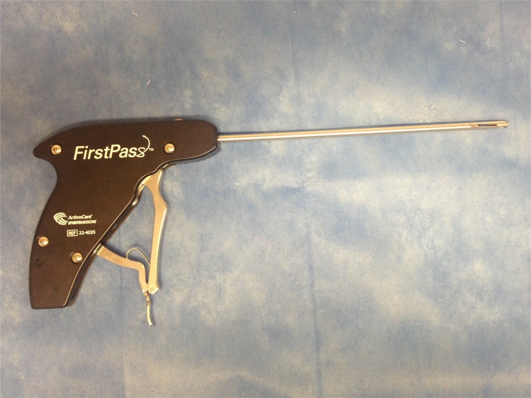
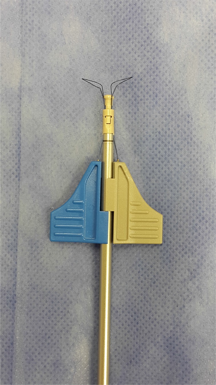
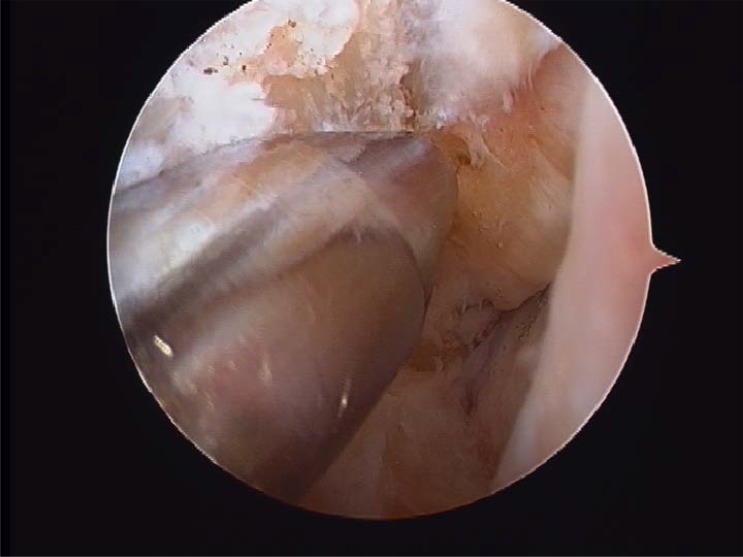
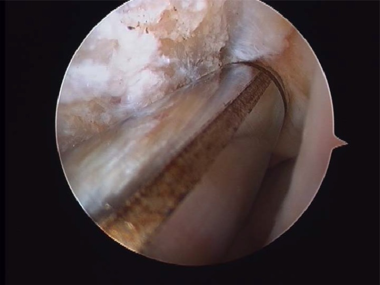
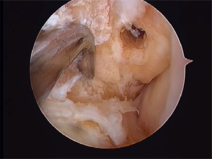
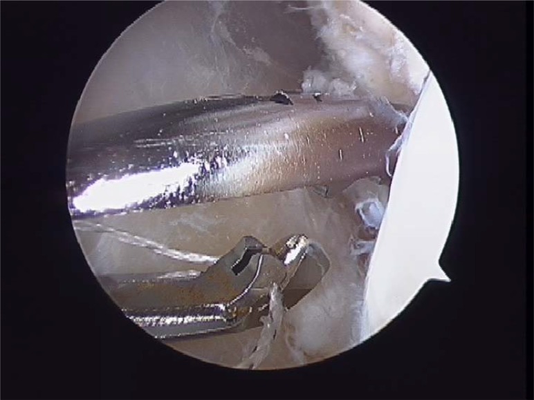
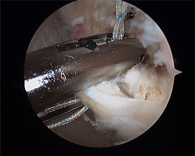
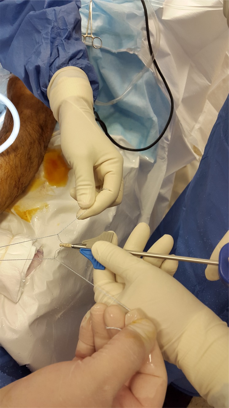
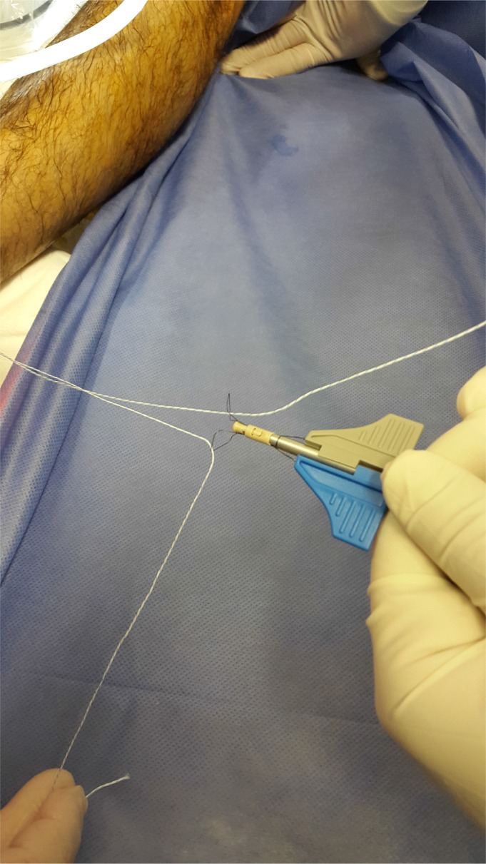
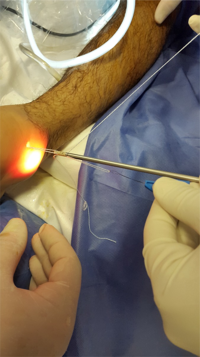
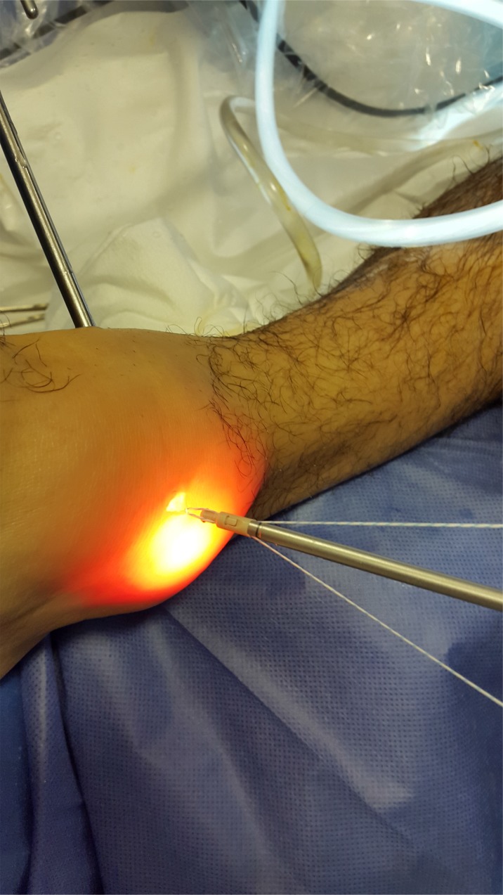
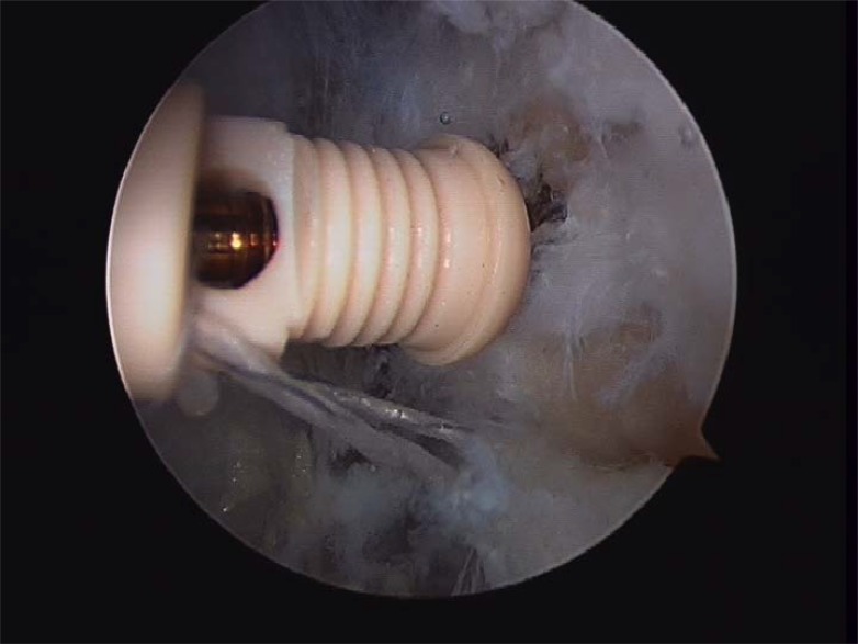
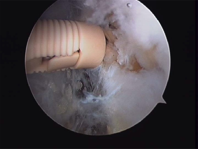
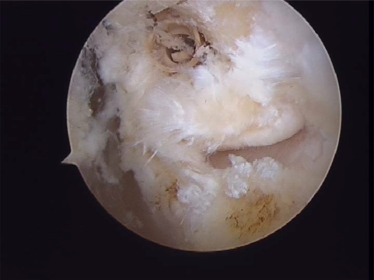
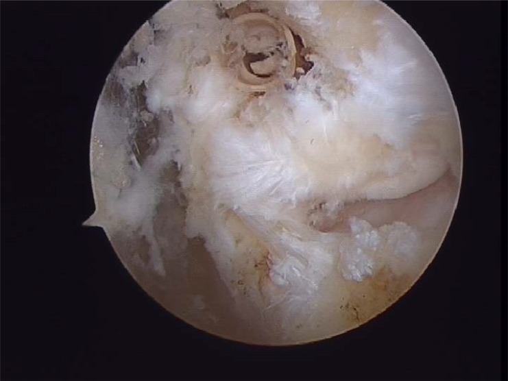
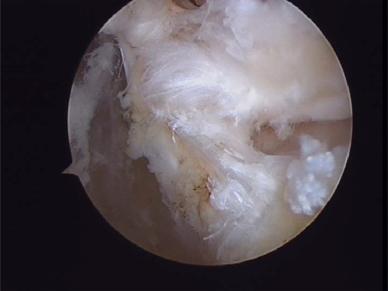
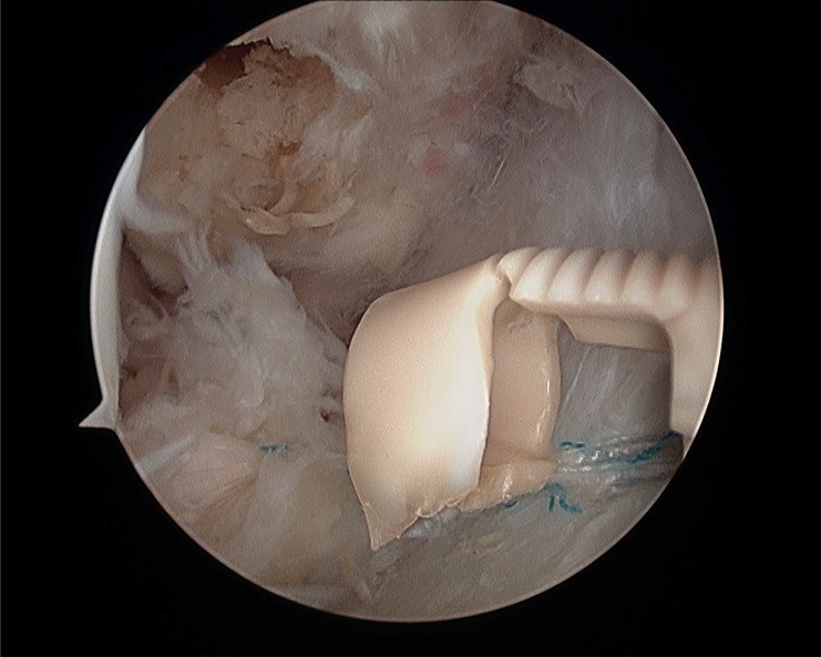
Material and methods: Of the 22 patients who were treated; 18 males and 4 females, and aged from 17-42 years (mean 28 years). All patients presented a history of more than three ankle sprains in the last two years and presented positive anterior drawer and talar tilt test of the ankle in the physical examination. We perform an anterior arthroscopy of the ankle in order to treat asociated disease and then we performed "All inside¨ lateral ligament repair through two portals (anteromedial and anterolateral) using an anchor knotless suture.

Results: Clinical outcome evaluations were performed at a mean follow up of 25 months. (R: 17-31). Overall results has been shown by means of the American Orthopaedic Foot and Ankle Society (AOFAS). Mean AOFAS scores improved from 63 points (range 52-77) preoperatively to 90 points (range 73-100) at final follow up. No recurrences of ankle instability were found in the cases presented.

Conclusion: Several surgical procedures have been described during the last years in order to treat chronic ankle instability. ¨All inside¨ lateral ligament reconstruction presents lower local morbidity than open procedures with few complications. Moreover, it is a reproductible technique, with high clinical success rate, few complications and relatively quick return to sports activities. A high knowledge of the anatomic landmarks should be essential to avoid unwated injuries.

Keywords: Ankle; Arthroscopy; Chronic instability; Reconstruction.


Figures

Fig. (1)
Fig. (1)
Arthroscopic approach in dorsiflexion (Anteromedial and anterolateral portals).
Fig. (2)
Fig. (2)
Lateral gutter.
Fig. (3)
Fig. (3)
Tibial exostosis.
Fig. (4A)
Fig. (4A)
Anterior talofibular partial rupture.
Fig. (4B)
Fig. (4B)
Complete anterior talofibular rupture.
Fig. (5)
Fig. (5)
Fibular footprint ATFL.
Fig. (6A)
Fig. (6A)
First Pass Suture Passer.
Fig. (6B)
Fig. (6B)
Anchor knotless suture 4, 5mm.
Fig. (7A)
Fig. (7A)
Fig. (7B)
Fig. (7B)
Fig. (7C)
Fig. (7C)
(Sequence Pictures) Performing the hole in fibular´s footprints.
Fig. (8A)
Fig. (8A)
(Sequence Pictures) Grasping the ligament with a HIFI suture is the first step for the repair. (A).
Fig. (8B)
Fig. (8B)
The First Pass suture passer is introduced through the anterolateral portal and the ligament is penetrated from lateral to medial (B).
Fig. (8C)
Fig. (8C)
Pull back the suture to know if there is a good capture of the remanent tissue (C).
Fig. (9A)
Fig. (9A)
Fig. (9B)
Fig. (9B)
Introducing the sutures into de anchor knotless loop.
Fig. (10A)
Fig. (10A)
Fig. (10B)
Fig. (10B)
Introducing the anchor.
Fig. (11A)
Fig. (11A)
Impacting the anchor and performing tension through the suture and remanent tissue.
Fig. (11B)
Fig. (11B)
Impacting the anchor and performing tension through the suture and remanent tissue.
Fig. (12A)
Fig. (12A)
Final ligament repair . The ligament is attached on its footprints and can be seen from anterolateral portal.
Fig. (12B)
Fig. (12B)
Final ligament repair . The ligament is attached on its footprints and can be seen from anterolateral portal.
Fig. (12C)
Fig. (12C)
Final ligament repair . The ligament is attached on its footprints and can be seen from anterolateral portal.
Fig. (13)
Fig. (13)
Anchor rupture during the impactation.

References

    1. Ekstrand J., Tropp H. The incidence of ankle sprains in soccer. Foot Ankle. 1990;11(1):41–44. doi: 10.1177/107110079001100108. - DOI - PubMed
    1. Garrick J.G. The frequency of injury, mechanism of injury, and epidemiology of ankle sprains. Am. J. Sports Med. 1977;5(6):241–242. doi: 10.1177/036354657700500606. - DOI - PubMed
    1. Guillo S., Archbold P., Perera A., Bauer T., Sonnery-Cottet B. Arthroscopic anatomic reconstruction of the lateral ligaments of the ankle with gracilis autograft. Arthrosc. Tech. 2014;3(5):e593–e598. doi: 10.1016/j.eats.2014.06.018. - DOI - PMC - PubMed
    1. Bosien W.R., Staples O.S., Russell S.W. Residual disability following acute ankle sprains. J. Bone Joint Surg. Am. 1955;37-A(6):1237–1243. doi: 10.2106/00004623-195537060-00011. - DOI - PubMed
    1. Freeman M.A. Instability of the foot after injuries to the lateral ligament of the ankle. J. Bone Joint Surg. Br. 1965;47(4):669–677. - PubMed

Publication types

LinkOut - more resources