

Conventional Versus Accelerated Collagen Cross-Linking for Keratoconus: A Comparison of Visual, Refractive, Topographic and Biomechanical Outcomes
- PMID: 29081866
- PMCID: PMC5633701
- DOI: 10.2174/1874364101711010262
Conventional Versus Accelerated Collagen Cross-Linking for Keratoconus: A Comparison of Visual, Refractive, Topographic and Biomechanical Outcomes
Abstract
Objective: The aim was to compare the visual, refractive, topographic and biomechanical outcomes in patients with progressive keratoconus treated with either conventional or accelerated crosslinking at one year follow up.
Methods: It is a prospective, non-randomised interventional study of 76 patients who underwent conventional (CXL; 3mW/cm2 for 30 minutes) or accelerated cross linking (KXL; 30mW/cm2 for 4 minutes) for progressive keratoconus. Baseline and postoperative visual acuity, manifest refraction, corneal topography, pachymetry, endothelial cell density and biomechanical parameters of corneal hysteresis and corneal resistance factor were evaluated and compared.
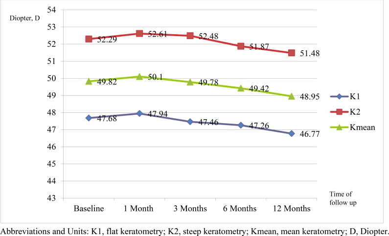
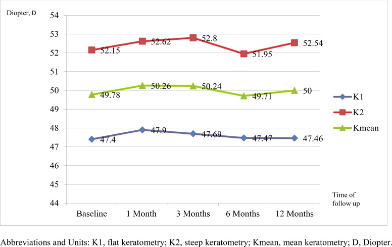
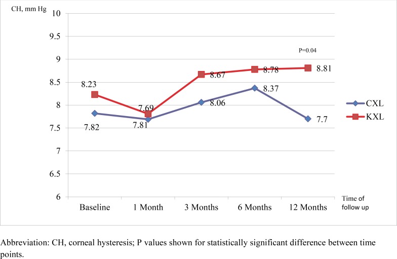
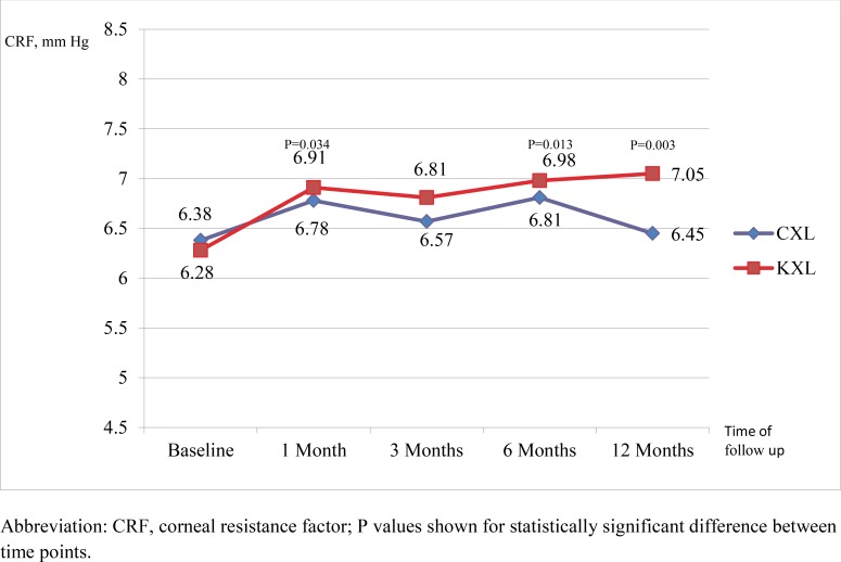
Results: The 2 groups were comparable in terms of uncorrected and best corrected visual acuity and spherical equivalent. Both groups showed no significant increase in K1, K2 and Kmean from baseline at 12 months. There was also no difference between the CXL and KXL group for postoperative corneal topography as well as central and minimal pachymetry up to 12 months. There was a significant increase in both corneal hysteresis (0.62mm Hg, P=0.04) and corneal resistance factor (0.91mm Hg, P=0.003) in the KXL group at 12 months but not in the CXL group. There was no significant endothelial cell loss throughout follow up in both the groups.
Conclusion: We have established comparability of the 2 protocols in stabilizing the progression of keratoconus. Our findings also suggested an added biomechanical advantage of accelerated crosslinking at 1 year follow up.
Keywords: Biomechanical outcomes; Collagen; Corneal biomechanics; Cross linking; Keratoconus; Topographic.
Figures
References
LinkOut - more resources
Full Text Sources
Other Literature Sources