. 2017 Sep 22;8(10):4640-4651.
doi: 10.1364/BOE.8.004640. eCollection 2017 Oct 1.

Non-invasive monitoring of skin inflammation using an oxygen-sensing paint-on bandage

Affiliations

Non-invasive monitoring of skin inflammation using an oxygen-sensing paint-on bandage

Zongxi Li et al. Biomed Opt Express. .

Abstract

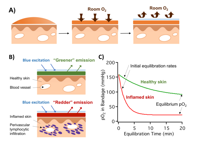
Inflammation involves a cascade of cellular and molecular mediators that ultimately lead to the infiltration of immune cells into the affected area. This inflammatory process in skin is common to many diseases including acne, infection, and psoriasis, with the presence or absence of immune cells a potential diagnostic marker. Here we show that skin inflammation can be non-invasively measured and mapped using a paint-on oxygen sensing bandage in an in vivo porcine inflammation model. After injection of a known inflammatory agent, the bandage could track the increase, plateau, and decrease in oxygen consumption at the injury site over 7 weeks, as well as discern inflammation resultant from injection at various depths beneath the surface of the skin. Both the initial rate of pO2 change and the change in bandage pO2 at equilibration (CBP20) were found to be directly related to the metabolic oxygen consumption rate of the tissue in contact. Healthy skin demonstrated an initial pO2 decrease rate of 6.5 [Formula: see text], and CBP20 of 84 [Formula: see text]. Inflamed skin had a significantly higher initial consumption rate of 55 [Formula: see text], and a larger CBP20 of 140 [Formula: see text]. The change in the bandage pO2 before and after equilibration with tissue was found to correlate well with histological evidence of skin inflammation in the animals.

Keywords: (160.2540) Fluorescent and luminescent materials; (170.2655) Functional monitoring and imaging; (170.3880) Medical and biological imaging; (170.6510) Spectroscopy, tissue diagnostics.


Figures

Fig. 1
Fig. 1
A) Illustration of the oxygen-sensing paint-on bandage applied to skin as a liquid, drying into a solid thin film, and the application of the barrier layer. B) Top: bandage applied to healthy skin; bottom: bandage applied to inflamed skin with perivascular infiltrating lymphocytes actively consuming oxygen. C) Upon application, bandage pO2 equilibrates with the tissue underneath: higher equilibrium pO2 and a slower initial equilibration rate when applied to healthy skin; lower equilibrium pO2 and a faster initial equilibration rate when applied to inflamed skin.
Fig. 2
Fig. 2
Tracking the progression of tissue inflammation in skin following intradermal CFA injection using the oxygen-sensing paint-on bandage. A) initial equilibration rate, calculated as the average pO2 change in the bandage within the first 4 minutes of bandage application, changes over the course of 7 weeks after CFA injection. B) CBP20 calculated as the difference between the initial and equilibrium oxygen partial pressure within the sensing bandage, tracked over 7 weeks post-injection. C) Correlating bandage equilibrium pO2 with the degree of tissue inflammation assessed by histological analyses. Top: equilibrium pO2 map captured by the oxygen-sensing bandage for skin with no CFA injection, 2nd week and 4th week after CFA injection. Equilibrium pO2 drops significantly after 2 weeks of injection, and returns after 4 weeks. Arrows pointing to outer square of the injection area marked by ink (also seen in the regular photograph); injection site is 0.5 inch to the right side of the line. Middle: regular photographs of skin with no CFA injection, 2nd week and 4th week after CFA injection. Bottom: histology of skin no CFA injection, 2nd week and 4th week after CFA injection. Focal epidermal and dermal inflammation was observed after 2 weeks of injection, which subsides after 4 weeks. Arrows pointing to perivascular inflammatory infiltrate.
Fig. 3
Fig. 3
A) Illustration of the different subcutaneous injection depths used in the experiment. B) CBP20 measured by the oxygen-sensing bandage on day 9 after CFA injection at the different injection depths. C) Confirming the degree of tissue inflammation by histological analysis. From top to bottom: regular photographs, histology (epiderm/derm), histology (derm/subcutaneous) at injection sites I, II, III and IV.

References

    1. Pasparakis M., Haase I., Nestle F. O., “Mechanisms regulating skin immunity and inflammation,” Nat. Rev. Immunol. 14(5), 289–301 (2014). 10.1038/nri3646 - DOI - PubMed
    1. Lowes M. A., Suárez-Fariñas M., Krueger J. G., “Immunology of psoriasis,” Annu. Rev. Immunol. 32(1), 227–255 (2014). 10.1146/annurev-immunol-032713-120225 - DOI - PMC - PubMed
    1. Buchman A. L., “Side effects of corticosteroid therapy,” J. Clin. Gastroenterol. 33(4), 289–294 (2001). 10.1097/00004836-200110000-00006 - DOI - PubMed
    1. Sina B., Kao G. F., Deng A. C., Gaspari A. A., “Skin biopsy for inflammatory and common neoplastic skin diseases: optimum time, best location and preferred techniques. A critical review,” J. Cutan. Pathol. 36(5), 505–510 (2009). 10.1111/j.1600-0560.2008.01175.x - DOI - PubMed
    1. Yoshinaga Y., Higaki M., Terajima S., Ohkubo E., Nogita T., Miyasaka N., Kawashima M., “Detection of inflammatory cytokines in psoriatic skin,” Arch. Dermatol. Res. 287(2), 158–164 (1995). 10.1007/BF01262325 - DOI - PubMed

LinkOut - more resources