. 2017 Dec 1;127(12):4326-4337.
doi: 10.1172/JCI94417. Epub 2017 Oct 30.

GLIS3 is indispensable for TSH/TSHR-dependent thyroid hormone biosynthesis and follicular cell proliferation

Affiliations

GLIS3 is indispensable for TSH/TSHR-dependent thyroid hormone biosynthesis and follicular cell proliferation

Hong Soon Kang et al. J Clin Invest. .

Abstract

Deficiency in Krüppel-like zinc finger transcription factor GLI-similar 3 (GLIS3) in humans is associated with the development of congenital hypothyroidism. However, the functions of GLIS3 in the thyroid gland and the mechanism by which GLIS3 dysfunction causes hypothyroidism are unknown. In the current study, we demonstrate that GLIS3 acts downstream of thyroid-stimulating hormone (TSH) and TSH receptor (TSHR) and is indispensable for TSH/TSHR-mediated proliferation of thyroid follicular cells and biosynthesis of thyroid hormone. Using ChIP-Seq and promoter analysis, we demonstrate that GLIS3 is critical for the transcriptional activation of several genes required for thyroid hormone biosynthesis, including the iodide transporters Nis and Pds, both of which showed enhanced GLIS3 binding at their promoters. The repression of cell proliferation of GLIS3-deficient thyroid follicular cells was due to the inhibition of TSH-mediated activation of the mTOR complex 1/ribosomal protein S6 (mTORC1/RPS6) pathway as well as the reduced expression of several cell division-related genes regulated directly by GLIS3. Consequently, GLIS3 deficiency in a murine model prevented the development of goiter as well as the induction of inflammatory and fibrotic genes during chronic elevation of circulating TSH. Our study identifies GLIS3 as a key regulator of TSH/TSHR-mediated thyroid hormone biosynthesis and proliferation of thyroid follicular cells and uncovers a mechanism by which GLIS3 deficiency causes neonatal hypothyroidism and prevents goiter development.

Keywords: Endocrinology; Thyroid disease.


Conflict of interest statement

Conflict of interest: The authors have declared that no conflict of interest exists.

Figures

Figure 1
Figure 1. TH levels are reduced and TSH levels elevated in Glis3KO mice.
(A) Comparison of serum T3, T4, and TSH levels between 1-month-old male and female WT (MWT and FWT, respectively) and Glis3KO (MKO and FKO, respectively) mice (n = 7–16). (B) The content of free T3, free T4, and thyroglobulin-bound T3 and T4 (Tg T3 and Tg T4, respectively) were analyzed in thyroid tissues from 1-month-old male and female WT and Glis3KO mice (n = 4). The content of hormones was normalized to tissue weight. Data are shown as mean ± SEM. (C) Glis3 is highly expressed in the thyroid. Levels of Glis3 mRNA in the liver, thyroid gland, pancreatic islets, and P7 testis were analyzed by QRT-PCR. Levels of expression were examined in tissues from at least 2 different mice. (D) Sections of thyroids from 1-week-old Glis3-EGFP mice were examined by immunohistochemistry with anti-GFP and anti-PECAM1 antibodies as described in Methods. Scale bar: 50 μm. (E) Thyroid tissues from 1-, 2-, and 4-week-old Glis3-EGFP mice were examined with antibodies against GFP and PAX8. Scale bar: 50 μm. *P < 0.05; **P < 0.001, Student’s t test..
Figure 2
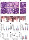
Figure 2. GLIS3 deficiency inhibits thyroid follicular cell proliferation and prevents goiter development in mice fed a LID.
(A) Thyroid glands from 1-, 2-, and 4-week-old WT and Glis3KO mice and (B) WT-LID and KO-LID mice were examined by H&E staining. Scale bars: 30 μm. (C) Representative thyroid glands with trachea from 4-week-old WT and Glis3KO mice and WT-LID and KO-LID mice. (DF) Ratios of the surface area of thyroid follicles over total surface area of the thyroid (D), the average size of the follicles (E), and the average number of follicular cells per follicle (F) were analyzed in thyroid glands from 1-, 2-, and 4-week-old WT and Glis3KO mice. n ≥ 2 for each group. (G and H) WT and Glis3KO mice were fed a LID for 6 days before serum TSH levels (G) and the relative weight of the thyroids (H) were determined. (I) Thyroid follicular cells from KO-LID mice did not become hypertrophic. Comparison of the average thyroid follicular cell size between WT and Glis3KO mice. n ≥ 2 for each group. Data are shown as mean ± SEM. *P < 0.05; **P < 0.001; ***P < 0.0001, Student’s t test.
Figure 3
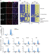
Figure 3. Proliferation of thyroid follicular cells was greatly reduced in KO-LID mice.
(A) EdU incorporation was analyzed in the thyroid of 3-week-old WT and Glis3KO mice and WT-LID and KO-LID mice as described in Methods. Arrows indicate PAX8+EdU+ cells, and arrowheads indicate PAX8EdU+ cells. Scale bar: 50 μm. (B) Percentages of PAX8+ cells staining EdU+ were calculated and plotted. n ≥ 3 for each group. (C) Heatmaps generated from gene expression profiles obtained by microarray analyses of thyroid glands from WT and Glis3KO mice fed either ND or LID as indicated. The same genes associated with cell cycle, TH biosynthesis, ECM, and cytokine/chemokine pathways as shown in Supplemental Table 3 are clustered as indicated. (D) QRT-PCR analysis of the expression of several cell proliferation regulatory genes in WT and Glis3KO mice fed with ND or LID. n ≥ 4. Data are shown as mean ± SEM. *P < 0.05; **P < 0.001; ***P < 0.0001, Student’s t test.
Figure 4
Figure 4. TSH/TSHR-mediated activation of the mTORC1 signaling pathway is impaired in the thyroid gland of KO-LID mice.
(A) Protein lysates obtained from thyroid glands of WT and Glis3KO mice fed either ND or LID were examined by Western blot analysis using RPS6, p-RPS6, and p-AKTS473 antibodies. (B) The relative levels of expression were analyzed and the ratio of p-RPS6/RPS6 plotted. n = 4 for each group. Data are shown as mean ± SEM. ***P < 0.0001, Student’s t test. (C) Representative images of sections of WT and Glis3KO thyroid tissue immunostained with antibodies against p-RPS6 (green), WGA (red), and DAPI. Scale bar: 50 μm.
Figure 5
Figure 5. Loss of GLIS3 function decreases the expression of several genes required for TH biosynthesis.
(A) Relative gene expression in thyroids from WT and Glis3KO mice fed either ND or LID was analyzed by QRT-PCR analysis. n ≥ 4 for each group. (B) Protein lysates obtained from thyroids of WT and Glis3KO mice fed either ND or LID were examined by Western blot analysis with antibodies against NIS and PAX8. GAPDH was used as a loading control. (C) Representative images of sections of WT and Glis3KO thyroids immunoassayed with antibodies against NIS (green), WGA (red), and DAPI. Scale bar: 50 μm. (D) The relative intensity of NIS immunostaining was analyzed with ImageJ (NIH) and plotted. n ≥ 2 for each group. (E) PCCl3-pIND20-Glis3 cells stably expressing Glis3 under the control of a Dox-inducible promoter were treated with or without Dox in the presence or absence of TSH and insulin (Ins). The expression of Glis3 and Nis was examined by QRT-PCR. (F) PCCl3 cells stably expressing Glis3 shRNA under the control of a Dox-inducible promoter to knock down Glis3 expression were treated with or without Dox in the presence or absence of TSH and insulin. The expression of Glis3 and Nis was examined by QRT-PCR. Data in E and F are derived from triplicate samples and representative of 2 independent experiments. Data are shown as mean ± SEM. *P < 0.05; **P < 0.001; ***P < 0.0001, Student’s t test. rGlis3 and rNis refer to rat genes.
Figure 6
Figure 6. GLIS3 regulates the transcription of a subset of differentially expressed genes directly through its interaction with GLIS3-BS.
(A) Pie chart showing the position of GLIS3-binding regions on the genome of the mouse thyroid gland relative to their nearest gene identified by Chipset analysis. The promoter is defined as the region up to 5 kb upstream of the TSS. (B) De novo consensus motif analysis using MEME program identified a G-rich GLIS3-binding motif, which is similar to GLIS3-BS identified previously by an in vitro binding assay. (C) GLIS3-EGFP occupancy on genome loci of several GLIS3-regulated genes associated with TH biosynthesis or cell proliferation. Tshr and Ccnb1 are included as negative controls. Gene tracks were taken from the UCSC Genome Browser (https://genome.ucsc.edu/). For each locus, Chipset analyses of input chromatin (Input) chromatin pulled down with GFP-BP-agarose (GFP-ChIP) from GLIS3-EGFP and WT thyroid glands are shown as indicated. GLIS3-binding peaks were observed only in IP GFP. (D) Schematic view of the key regulatory functions of GLIS3 in thyroid follicular cells. TSH/TSHR activates several kinase pathways that subsequently lead to increased transcription of many genes required for TH biosynthesis. GLIS3 is essential for TH biosynthesis, acting by directly regulating the transcription of several key TH biosynthetic genes, particularly Pds and Nis. Prolonged elevated levels of TSH induce thyroid follicular cell proliferation and goiter development through AKT1-independent activation of the mTORC1 signaling pathway. GLIS3 is required for the activation of the mTORC1 pathway and thyroid follicular cell proliferation. GLIS3 is critical in mediating the downstream actions of TSH/TSHR. GLIS3 activity might be controlled by TSH/TSHR and regulated by one of the TSH/TSHR-activated kinase pathways.

References

    1. Mondal S, Raja K, Schweizer U, Mugesh G. Chemistry and biology in the biosynthesis and action of thyroid hormones. Angew Chem Int Ed Engl. 2016;55(27):7606–7630. doi: 10.1002/anie.201601116. - DOI - PubMed
    1. Kleinau G, Neumann S, Grüters A, Krude H, Biebermann H. Novel insights on thyroid-stimulating hormone receptor signal transduction. Endocr Rev. 2013;34(5):691–724. doi: 10.1210/er.2012-1072. - DOI - PMC - PubMed
    1. Bagriacik EU, Yaman M, Haznedar R, Sucak G, Delibasi T. TSH-induced gene expression involves regulation of self-renewal and differentiation-related genes in human bone marrow-derived mesenchymal stem cells. J Endocrinol. 2012;212(2):169–178. doi: 10.1530/JOE-11-0404. - DOI - PubMed
    1. Brewer C, Yeager N, Di Cristofano A. Thyroid-stimulating hormone initiated proliferative signals converge in vivo on the mTOR kinase without activating AKT. Cancer Res. 2007;67(17):8002–8006. doi: 10.1158/0008-5472.CAN-07-2471. - DOI - PubMed
    1. Dremier S, et al. The role of cyclic AMP and its effect on protein kinase A in the mitogenic action of thyrotropin on the thyroid cell. Ann N Y Acad Sci. 2002;968:106–121. doi: 10.1111/j.1749-6632.2002.tb04330.x. - DOI - PubMed

Publication types

MeSH terms