. 2017 Oct 30;12(10):e0186888.
doi: 10.1371/journal.pone.0186888. eCollection 2017.

TrpA1 activation in peripheral sensory neurons underlies the ionic basis of pain hypersensitivity in response to vinca alkaloids

Affiliations

TrpA1 activation in peripheral sensory neurons underlies the ionic basis of pain hypersensitivity in response to vinca alkaloids

Nina Boiko et al. PLoS One. .

Abstract

Chemotherapy induced peripheral neuropathy (CIPN), a side effect of many anti-cancer drugs including the vinca alkaloids, is characterized by a severe pain syndrome that compromises treatment in many patients. Currently there are no effective treatments for this pain syndrome except for the reduction of anti-cancer drug dose. Existing data supports the model that the pain associated with CIPN is the result of anti-cancer drugs augmenting the function of the peripheral sensory nociceptors but the cellular mechanisms underlying the effects of anti-cancer drugs on sensory neuron function are not well described. Studies from animal models have suggested a number of disease etiologies including mitotoxicity, axonal degeneration, immune signaling, and reduced sensory innervations but these outcomes are the result of prolonged treatment paradigms and do not necessarily represent the early formative events associated with CIPN. Here we show that acute exposure to vinca alkaloids results in an immediate pain syndrome in both flies and mice. Furthermore, we demonstrate that exposure of isolated sensory neurons to vinca alkaloids results in the generation of an inward sodium current capable of depolarizing these neurons to threshold resulting in neuronal firing. These neuronal effects of vinca alkaloids require the transient receptor potential ankyrin-1 (TrpA1) channel, and the hypersensitization to painful stimuli in response to the acute exposure to vinca alkaloids is reduced in TrpA1 mutant flies and mice. These findings demonstrate the direct excitation of sensory neurons by CIPN-causing chemotherapy drugs, and identify TrpA1 as an important target during the pathogenesis of CIPN.


Conflict of interest statement

Competing Interests: The authors have declared that no competing interests exist.

Figures

Fig 1
Fig 1. Vinca alkaloids generate neuropathic pain in Drosophila larvae.
A, Nocifensive stimuli to larvae elicit a stereotypical rolling response. B, Force-response curve for indicated forces. Graphs represent the average values for responding larvae. C-E, Increased responses of larvae to mechanical stimulation (50-60mN) after feeding vinblastine (C) or vincristine (D) at indicated concentrations for 1 hr (black bars) or 24 hrs (gray bars) versus vehicle controls. (E) Increased responsiveness persists after larvae are removed from drug. In all drug treatment studies, average treatment values were normalized to vehicle controls assayed in parallel. Gray line indicates vehicle (100%) values. F, Temperature-response curve for indicated temperatures. Graphs represent the average values for responding larvae. G, Increased response of larvae to thermal stimulation (37°C) after 1 hour feeding of vinblastine (50 μM). Graphs represent the average values for responding larvae. Error bars = SEM, n = 160–200 larvae assayed in groups of 20 per each condition. Significance determined by ANOVA except for drug treatments where significance was determined using a Student’s T-test for drug vs. vehicle for each condition except where indicated (*p<0.05). † indicate significant differences determined by ANOVA analysis (p<0.01).
Fig 2
Fig 2. Acute exposure to vinca alkaloids has no effect on C4da sensory neuron morphology or mitochondrial toxicity.
A, Confocal images of dendritic arbors, axonal projections (i), and axonal nerve terminals (ii) from C4da neurons in larvae fed vinblastine (10 μM) or vehicle for 1 hr prior to imaging. B-C, Morphometric analysis reveals no significant effects of vinblastine (black bars) on total dendritic length (A), dendritic crossing (B), and axonal terminal length (C) compared to C4da neurons incubated with vehicle (gray bars). Bars represent average values. Error bars = SEM, n = 10 dendrites; 17–20 nerve terminals. E, Representative recording showing the calcium overload required to induce mPTP opening (arrow) in isolated mitochondria. F, Bar graph showing no significant differences in the calcium retention capacity (CRC) of isolated mitochondria treated with 10 uM vinblastine (black bar) compared to vehicle (gray bar). Bars represent average values. Error bars = SEM, n = 7 animals for each condition.
Fig 3
Fig 3. Vinca alkaloids increase the excitability of C4da sensory neurons.
A-C, Representative traces from current-clamped C4da neurons show that exposure to vinblastine (A, B) or vincristine (C) immediately depolarizes the neuron and generates action potentials that is sensitive to the dose and presence of the drug. D and E, Current injections determine the relationship between current and firing frequency in C4da neurons (D). These analyses reveal that the firing frequencies resulting from vinca alkaloids (E) are consistent with amplitudes of the resulting current measured in voltage-clamp. Data for vinblastine (1, 10, and 50 μM) are indicated in red. Points represent average values. Error bars = SEM, n = 8–15 neurons per condition. (F) Graph represents the relationship between current injection and action potential frequency in the presence (arrowhead) and absence (circle) of sub-threshold concentrations of vinblastine. Each point represents the average frequency. Error bars = SEM, n = 3–5 neurons per condition. *p<0.05 determined via Student’s T-test for vinblastine vs. control for each current injection.
Fig 4
Fig 4. Vinca alkaloids directly excite C4da sensory neurons.
A-B, Representative traces from voltage-clamped C4da neurons held at indicated voltages and challenged with 50 μM vinblastine (A) or 50 μM vincristine (B) reveal an immediate inward current that is sensitive to HC-030031 (A) and Ruthenium red (RR) (B). C, Graph represents the average values for the currents measured in voltage-clamped C4da sensory neurons resulting from the indicated treatments. * p<0.01 indicate values significantly different than all other values as determined by ANOVA. Error bars = SEM, n = 3–18 neurons per condition. D, Values represent the average current (I) obtained at each holding voltage (V) in C4da neurons exposed to vehicle (black line) or 50 uM vinblastine (gray line). *p<0.01 determined for each value by a Student’s T-test. Error bars = SEM, n = 3–4 neurons per condition. E, Graph represents the average nociceptive responses of larvae fed 50 uM Vinblastine in the absence (gray bar) or presence of 100 uM HC-030031 (blue bar). * p < 0.05 determined by Student’s T-test comparing each condition. Gray line indicates the value obtained for vehicle control larvae. Error bars = SEM, n = 160 larvae per condition. F-G, Graphs represent the average gene expression level of all transcripts of the indicated Drosophila TRP family genes in purified C4da neurons (F; gray bars) and in whole larvae (G; black bars). tpm = transcripts per million. Error bars = sem. n = 4 independent samples for each condition.
Fig 5
Fig 5. The effects of vinca alkaloids on neuron excitability are reduced in dTrpA1 mutant C4da sensory neurons.
A-D, Current-clamp recordings of C4da neurons reveal that the dosage effects of vinblastine on neuronal firing frequencies (A and D) are completely absent in dTrpA1 mutant C4da neurons (B and D, red bars). In contrast, pain mutant C4da respond robustly to vinblastine (C and D, gray bars). D, Graph represents average values for action potential frequencies as a function vinblastine dose from C4da sensory neurons isolated from wild type (wt, black bars), dTrpA1 (red bars), and pain (gray bars) mutants. * p<0.05, ** p<0.001 indicate values significantly different than all other values as determined by ANOVA. Error bars = SEM, n = 3–15 neurons per condition. E, Comparison of current injection in dTrpA1 mutant (B) to wild type finds no difference in the excitability of wild type (black line) and dTrpA1 (red line) C4da neurons.
Fig 6
Fig 6. The effects of vinblastine on sensory neurons are absent in dTrpA1 mutants.
A and B, Representative traces from voltage-clamped C4da sensory neurons isolated from dTrpA1 (A) or pain (B) mutants held at indicated voltages and challenged with 50 μM vinblastine. C, Graph represents the average values for the currents measured in voltage-clamped C4da sensory neurons from wild type (black bars; data from Fig 4C for comparison), dTrpA1 (red bar), and pain (gray) mutants incubated with 50 uM vinblastine with or without HC-030031 (+HC). * p<0.01 indicate significant difference determined by Student’s T-test. Error bars = SEM, n = 3–6 neurons per condition. D, Behavioral analysis of dTrpA1 (red bars) and pain (gray bars) mutant larvae find that the effects of vinblastine are specifically absent in dTrpA1 mutants and that re-expression of wild type dTrpA1 only in C4da neurons rescues this deficit in dTrpA1 mutants. For these genetic analyses, average values were normalized to the vehicle controls assayed in parallel of the same experimental genotype. Gray line indicates vehicle (100%) values. * p < 0.05 determined by Student’s T-test of drug vs. vehicle for each genotype. Error bars = SEM, n = 160–200 larvae per condition.
Fig 7
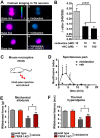
Fig 7. Acute exposure to vinblastine activates mammalian sensory neurons and generates pain that is reduced in TRPA1 knockout mice.
A Representative images of Trp-V1 expressing TG neurons challenged with vinblastine (10 uM, panel ii), (100 uM, panel iv), and vinblastine + HC-030031 (10 uM; panel vi). Base line fluorescence images shown in panels i, iii, and v. B Graphs represent the values for the average calcium accumulation within Trp-V1 expressing TG neurons challenged with 10 uM vinblastine (gray bar) or vinblastine + HC-030031 (black bars). Error bars = SEM. n = 23–32 neurons from 4 slides per condition. P value determined using ANOVA analysis. Control noise level indicated by gray line. C and D, Single injection of vinblastine (squares) directly into the hindpaw of wild type mice (C) results in increased spontaneous pain compared to vehicle (circles). Values represent average duration (in seconds) in bouts of nocifensive behavior. Error bars = SEM, n = 7 animals per group. *p<0.05 determined by Student’s T-test for each point versus vehicle control. E and F, Single injection of vinblastine (striped bars) into the hindpaw results in a significant mechanical allodynia (E) and thermal hyperalgesia (F) compared to control injections (solid bars). This effect is significantly reduced in TrpA1 knock-out mice (TRPA1 KO, red bars). ** p<0.01 determined by Student’s T-test comparing vinblastine versus control values. † p<0.01 determined by Student’s T-test comparing wild type versus TRPA1 mutants. Bars represent average values. Error bars = SEM, n = 7 animals per group.

References

    1. Malik B, Stillman M. Chemotherapy-induced peripheral neuropathy. Curr Neurol Neurosci Rep. Current Science Inc; 2008;8: 56–65. doi: 10.1007/s11910-008-0010-5 - DOI - PubMed
    1. Boyette-Davis JA, Walters ET, Dougherty PM. Mechanisms involved in the development of chemotherapy-induced neuropathy. Pain Management. 2015;5: 285–296. doi: 10.2217/pmt.15.19 - DOI - PMC - PubMed
    1. Jaggi AS, Singh N. Mechanisms in cancer-chemotherapeutic drugs-induced peripheral neuropathy. Toxicology. 2012;291: 1–9. doi: 10.1016/j.tox.2011.10.019 - DOI - PubMed
    1. Bhattacharya MRC, Gerdts J, Naylor SA, Royse EX, Ebstein SY, Sasaki Y, et al. A Model of Toxic Neuropathy in Drosophila Reveals a Role for MORN4 in Promoting Axonal Degeneration. Journal of Neuroscience. 2012;32: 5054–5061. doi: 10.1523/JNEUROSCI.4951-11.2012 - DOI - PMC - PubMed
    1. Nieto FR, Entrena JM, Cendán CM, Pozo ED, Vela JM, Baeyens JM. Tetrodotoxin inhibits the development and expression of neuropathic pain induced by paclitaxel in mice. Pain. 2008;137: 520–531. doi: 10.1016/j.pain.2007.10.012 - DOI - PubMed