. 2017 Dec 15;144(24):4625-4636.
doi: 10.1242/dev.154039. Epub 2017 Oct 30.

Conservation and variation in pair-rule gene expression and function in the intermediate-germ beetle Dermestes maculatus

Affiliations

Conservation and variation in pair-rule gene expression and function in the intermediate-germ beetle Dermestes maculatus

Jie Xiang et al. Development. .

Abstract

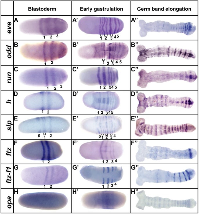
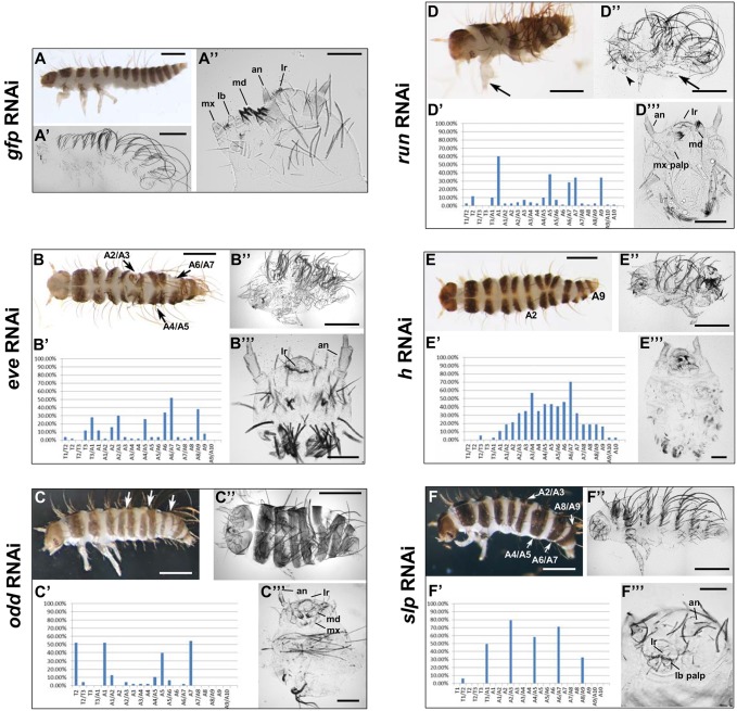
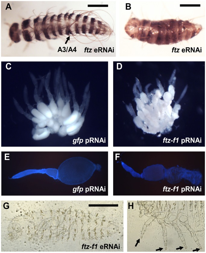
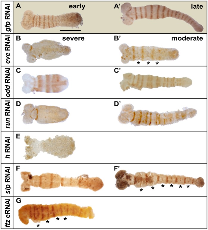
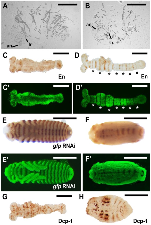
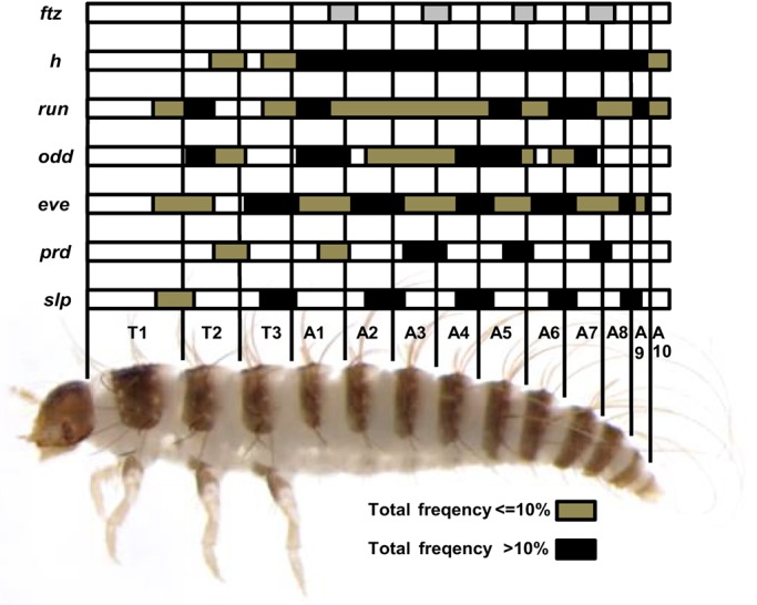
A set of pair-rule (PR) segmentation genes (PRGs) promotes the formation of alternate body segments in Drosophila melanogaster Whereas Drosophila embryos are long-germ, with segments specified more or less simultaneously, most insects add segments sequentially as the germband elongates. The hide beetle Dermestes maculatus represents an intermediate between short- and long-germ development, ideal for comparative study of PRGs. We show that eight of nine Drosophila PRG orthologs are expressed in stripes in Dermestes Functional results parse these genes into three groups: Dmac-eve, -odd and -run play roles in both germband elongation and PR patterning; Dmac-slp and -prd function exclusively as complementary, classic PRGs, supporting functional decoupling of elongation and segment formation; and orthologs of ftz, ftz-f1, h and opa show more variable function in Dermestes and other species. While extensive cell death generally prefigured Dermestes PRG RNAi-mediated cuticle defects, an organized region with high mitotic activity near the margin of the segment addition zone is likely to have contributed to truncation of eveRNAi embryos. Our results suggest general conservation of clock-like regulation of PR stripe addition in sequentially segmenting species while highlighting regulatory rewiring involving a subset of PRG orthologs.

Keywords: Beetle; Dermestes; Evo-devo; Germband elongation; Pair-rule gene; Segmentation.


Conflict of interest statement

Competing interestsThe authors declare no competing or financial interests.

Figures

Fig. 1.
Fig. 1.
Eight Dermestes PRG orthologs are expressed in PR stripes. Expression of Dermestes PRGs as assessed by in situ hybridization during early development at the stages indicated. Early gastrulation was defined by appearance of the ventral furrow. Note that Dermestes embryos vary in shape and size. (A-A″) Dmac-eve. (A) The first primary stripe has refined as a transverse stripe in the embryo's center. The second primary stripe is fuzzy and broad. A third stripe is resolving from cap-like expression in the posterior end. (A′) Five primary stripes have emerged. The anterior three have split to form secondary stripes (brackets). (A″) Stripes are fading in anterior segments but remain strong in posterior segments. (B-B″) Dmac-odd. (B) Two primary stripes are clear, along with cap-like expression in the posterior. (B′) Five primary stripes and three intercalated secondary stripes have emerged. (B″) Primary and secondary stripes alternate in intensity, with fading anterior expression. (C-C″) Dmac-run. (C) Three stripes have arisen with weak cap-like expression in the posterior end. (C′) In total, five stripes have emerged; the first and second are broader. (C″) Anterior expression has faded; weak stripes remain at the posterior end. (D-D″) Dmac-h. (D) Two Dmac-h stripes in the center of the embryo. (D′) At least five primary stripes are detectable. The second primary stripe has split into two thin stripes. (D″) Strong stripes in the most posterior two segments with faint expression in the anterior. (E-E″) Dmac-slp. (E) Three primary stripes with secondary stripes emerging for stripe 1 (bracket). The most anterior stripe (stripe 0) marks the future antennae. (E′) Stripe 0, four primary stripes and three secondary stripes are present. (E″) Segmental stripes show equal intensity, with fuzzy anterior borders. (F-F″) Dmac-ftz. (F) Two strong, central stripes. (F′) Four stripes at early gastrulation. (F″) Strong expression remains in the most posterior segment. (G-G″) Dmac-ftz-f1. (G) Lateral expression in two stripes. (G′) Four stripes are visible but Dmac-ftz-f1 is not expressed in the ventral furrow. (G″) Fading expression in the anterior; strong stripes in the posterior. (H-H″) Dmac-opa. (H) Uniform, weak expression at blastoderm. (H′) Ubiquitous expression in the trunk. (H″) Weak segmental stripes throughout the germband. Embryos are oriented with anterior, left.
Fig. 2.
Fig. 2.
Truncated embryos and PR defects after Dermestes PRG ortholog knockdown. Offspring of pRNAi for control gfp or for the indicated D. maculatus PRG orthologs. Bar charts indicate the frequency of defects (y-axis) in anterior to posterior segments (x-axis) of affected hatched larvae. Data were from hatched larvae with segmentation defects from three consecutive daily collections after 3′ dsRNA knockdowns. The patterns were very similar after 5′ dsRNA knockdowns. (A,A′) Control Dermestes larvae have three thoracic segments and ten abdominal segments. (A) Hatched larva with pigmented stripes on the head and every trunk segment. (A′) Cuticular preparation of unhatched larva without pigmentation. (A″) Head close-up shows antennae (an), labrum (lr), mandibles (md), maxillae (mx) and labium (lb). (B-B″) Dmac-evepRNAi. (B) Moderately affected hatched larva with multiple fused segments (arrows). (B′) T3/A1, A2/A3, A4/A5, A6/A7, A8/A9, and neighboring regions were most frequently affected (n=50). (B″) Unhatched larva with shortened body length. (B‴) Spherical cuticle of a severely affected unhatched larva consists of only head, with antenna and labrum. (C-C‴) Dmac-oddpRNAi. (C) Hatched larva with only two pairs of legs and several partially fused segments (arrows). (C′) T2, A1, A5 and A7 were most commonly affected (n=48). (C″) Unhatched larva with short body and fewer segments. (C‴) Severely affected unhatched offspring with head and some thoracic structure. (D-D‴) Dmac-runpRNAi. (D) Hatched larva has fewer segments and deformed leg with duplicated claw (arrow). (D′) T2, A1, A5, A7, A9, and adjacent regions most frequently affected (n=70). (D″) Unhatched, shortened larva with duplicated claw (arrow) and leg-like maxilla (arrowhead). (D‴) ‘Head-only’ cuticle lacking partial maxillary and labium structures. (E-E‴) Dmac-hpRNAi. (E) Hatched larva with irregular segmentation between A2 and A9. (E′) Regions between A1 and A9 most frequently affected (n=37). (E″,E‴) Unhatched larva with head and thoracic segments, but little or no segmented abdomen. No wild-type-like terminal structures are evident. (F-F‴) Dmac-slppRNAi. (F) Hatched larva with several fused adjacent segments (arrows). (F′) T3/A1, A2/A3, A4/A5, A6/A7 and A8/A9 are most frequently affected (n=101). (F″) Unhatched larvae with fewer trunk segments and only one pair of legs. (F‴) Unhatched larval head missing mandibles and maxillae. Scale bars: 500 µm, except 100 µm for A″,B‴,C‴,D‴,E‴,F‴.
Fig. 3.
Fig. 3.
Dmac-ftz and ftz-f1 RNAi. (A,B) Hatched offspring after Dmac-ftz eRNAi. (A) Mildly affected embryo with single segmental fusion (arrow). (B) Severely affected larva with shortened body length and fewer segments. (C,E) Wild-type-like ovaries from gfp dsRNA-injected control female. (C) Large, oval-shaped, developing oocyte in each ovariole. (E) DAPI staining of dissected ovariole reveals large oocyte. (D,F) Ovaries from Dmac-ftz-f1 dsRNA-injected females. (D) Small primary oocytes clustered in each ovariole. No mature oocyte is visible. (F) Dissected ovariole with shrunken oocyte. (G) Unhatched larva from Dmac-ftz-f1 dsRNA-injected female has normal number of segments without any obvious segmentation defect. (H) Arrows indicate truncated distal ends of legs. Scale bars: 500 µm.
Fig. 4.
Fig. 4.
En expression is altered after Dermestes PRG knockdown. (A,A′) Control embryo (from gfp dsRNA-injected female) shows En stripes with equal intensity in every segment at early or late germband elongation stages. (B) Dmac-evepRNAi severely affected embryo with truncated, asegmental germband only has antennal En expression. (B′) A less severely affected embryo shows reduced En in alternate stripes (asterisks). (C) Dmac-oddpRNAi severely affected embryo with truncated germband has enlarged En stripes. (C′) ‘Paired’ En stripes detected in a moderately affected embryo. (D) Dmac-runpRNAi severely affected embryo. En is only detected in antennal and mandibular segments. (D′) Fused En stripes are visible in moderately affected offspring. (E) Dmac-hpRNAi. No striped En expression pattern is detectable. (F,F′) Dmac-slppRNAi. Alternate En stripes are absent (F) or reduced (F′, asterisks). (G) Dmac-ftzeRNAi. Alternate En stripes are weak or absent (asterisks). Scale bar: 500 µm.
Fig. 5.
Fig. 5.
Dmac-prd and -slp double RNAi produces asegmental embryos. (A,B) Significantly shortened unhatched larvae after Dmac-prd, -slp double pRNAi. Only antenna and labrum are visible. No gnathal appendages or legs are present. (C) No striped-like En expression is detected in elongated germband in a severely affected embryo. (D) Moderately affected embryo shows reduced En expression. Reduction is more obvious for even-numbered En stripes (asterisks). (C′,D′) SYTOX Green staining of embryos in C and D for visualization of embryo morphology. Note that there are no obvious segmental grooves (C′) or fused adjacent segments (D′), indicating overt lack of segmentation or milder segmentation defects, respectively. (E) Striped En expression at the posterior border of each segment in control embryos. (F) Only residual En expression is detected along the midline after double knockdown. (E′,F′) SYTOX Green staining of embryos in E and F for visualization of embryo morphology. (G,H) Dcp-1 antibody staining for apoptosis at elongated germband stage (G) and in a later stage embryo (H). There is almost uniform apoptosis signal in the elongated germband (G). Note that we previously observed stronger effects for Dmac-prd using eRNAi than pRNAi (Xiang et al., 2015). Scale bars: 500 µm.
Fig. 6.
Fig. 6.
Increased apoptosis and disrupted mitosis after Dermestes PRG knockdown. (A-F) Dcp-1 (top) and SYTOX Green nuclear staining (bottom) in pRNAi offspring. (A) gfpRNAi control. Apoptosis is detected in the head and posterior at low levels after germband elongation. Primordial appendages are visible. (B) Dmac-evepRNAi. Increased apoptosis is seen in the trunk. Antennae primordia, but no appendages, are visible. (C) Dmac-oddpRNAi. Increased apoptosis in the trunk of the early embryo. Antennal, mandible, maxillary and some thoracic primordia are present. (D) Dmac-runpRNAi. Extensive apoptosis in the center of the embryo; only antennal and mandible appendages are present. (E) Dmac-hpRNAi. High apoptotic activity at the posterior end. (F) Dmac-slppRNAi. Striped apoptosis concentrated in alternate compartments is apparent in an extended germband stage embryo. (G-O) PH3 staining to monitor mitosis. (G-J) Control gfppRNAi. (G) Mitotic cells are clustered in head lobes at early germband elongation. (H) Later, there is increased mitosis signal in the head and along the anterior and central trunk. In the posterior, mitotic cells are arranged in a stripe (arrow). (I) Concentrated mitotic activity persists in the posterior at late germband elongation (arrow). (J) High mitotic activity all along the embryo at later stage. (K-M) Dmac-slppRNAi. PH3 staining very similar to control. Concentrated mitosis is seen in the posterior (arrows in K,L). (N,O) Dmac-evepRNAi. (N) Embryo during germband elongation has fewer mitotic cells and without a clear striped-like arrangement in the posterior. (O) Later stage embryo with extensive mitotic activity throughout. Among the 200 0-1 day AEL embryos that were examined for every staining, over 25% developed to appropriate developmental stages for the examination of apoptosis or mitosis pattern. Scale bars: 500 µm.
Fig. 7.
Fig. 7.
Dermestes PRG orthologs are required for elongation and PR-like segmentation. Schematic representation of defective patterns after PRG RNAi. Results are based on defective patterns in affected hatched larvae after pRNAi, except for ftz (eRNAi, gray bars). Black bars indicate regions affected at high frequency (>10%); tan bars show regions affected at low frequency (≤10%). For genes playing dual roles in Dermestes segmentation (eve, odd, run), black bars alone show PR-like phenotypes in moderately affected offspring (hatched), whereas black and tan bars together indicate severely affected, truncated offspring (unhatched). PR-like defects seen for slppRNAi versus prdpRNAi or for ftzeRNAi versus evepRNAi are complementary.

Similar articles

Cited by

References

    1. Abzhanov A., Popadic A. and Kaufman T. C. (1999). Chelicerate Hox genes and the homology of arthropod segments. Evol. Dev. 1, 77-89. 10.1046/j.1525-142x.1999.99014.x - DOI - PubMed
    1. Akam M. (1987). The molecular basis for metameric pattern in the Drosophila embryo. Development 101, 1-22. - PubMed
    1. Angelini D. R., Liu P. Z., Hughes C. L. and Kaufman T. C. (2005). Hox gene function and interaction in the milkweed bug Oncopeltus fasciatus (Hemiptera). Dev. Biol. 287, 440-455. 10.1016/j.ydbio.2005.08.010 - DOI - PubMed
    1. Aranda M., Marques-Souza H., Bayer T. and Tautz D. (2008). The role of the segmentation gene hairy in Tribolium. Dev. Genes Evol. 218, 465-477. 10.1007/s00427-008-0240-1 - DOI - PMC - PubMed
    1. Auman T., Vreede B. M. I., Weiss A., Hester S. D., Williams T. A., Nagy L. M. and Chipman A. D. (2017). Dynamics of growth zone patterning in the milkweed bug Oncopeltus fasciatus. Development 144, 1896-1905. 10.1242/dev.142091 - DOI - PMC - PubMed

MeSH terms