. 2017 Oct 30;7(1):14293.
doi: 10.1038/s41598-017-14301-3.

Exosomal microRNA signatures in multiple sclerosis reflect disease status

Affiliations

Exosomal microRNA signatures in multiple sclerosis reflect disease status

Saeideh Ebrahimkhani et al. Sci Rep. .

Abstract

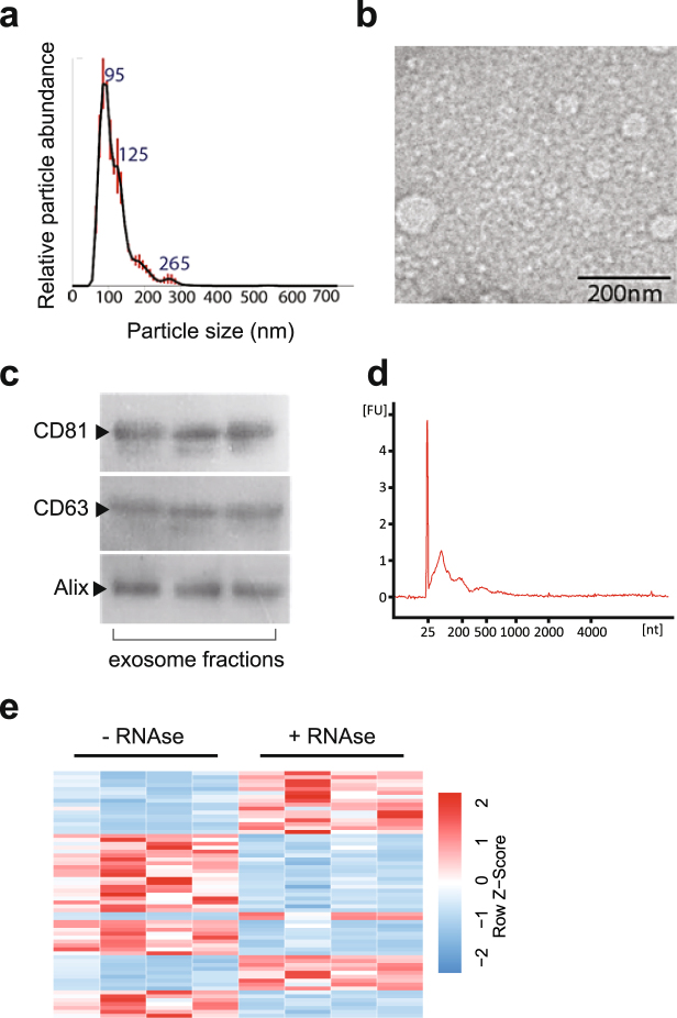
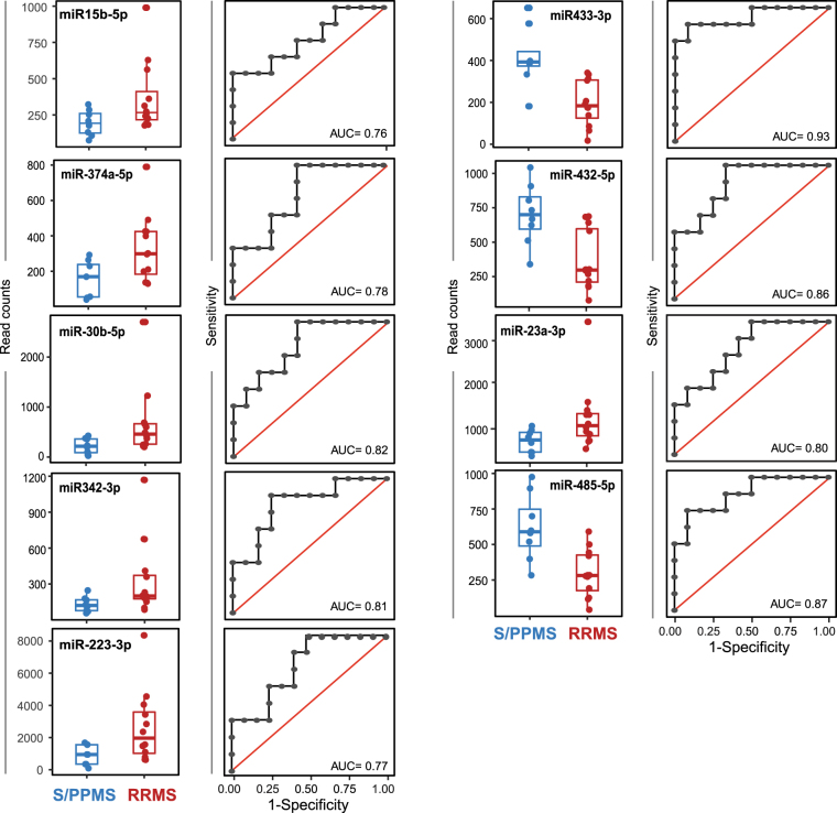
Multiple Sclerosis (MS) is a chronic inflammatory demyelinating disease of the central nervous system (CNS). There is currently no single definitive test for MS. Circulating exosomes represent promising candidate biomarkers for a host of human diseases. Exosomes contain RNA, DNA, and proteins, can cross the blood-brain barrier, and are secreted from almost all cell types including cells of the CNS. We hypothesized that serum exosomal miRNAs could present a useful blood-based assay for MS disease detection and monitoring. Exosome-associated microRNAs in serum samples from MS patients (n = 25) and matched healthy controls (n = 11) were profiled using small RNA next generation sequencing. We identified differentially expressed exosomal miRNAs in both relapsing-remitting MS (RRMS) (miR-15b-5p, miR-451a, miR-30b-5p, miR-342-3p) and progressive MS patient sera (miR-127-3p, miR-370-3p, miR-409-3p, miR-432-5p) in relation to controls. Critically, we identified a group of nine miRNAs (miR-15b-5p, miR-23a-3p, miR-223-3p, miR-374a-5p, miR-30b-5p, miR-433-3p, miR-485-3p, miR-342-3p, miR-432-5p) that distinguished relapsing-remitting from progressive disease. Eight out of nine miRNAs were validated in an independent group (n = 11) of progressive MS cases. This is the first demonstration that microRNAs associated with circulating exosomes are informative biomarkers not only for the diagnosis of MS, but in predicting disease subtype with a high degree of accuracy.


Conflict of interest statement

The authors declare that they have no competing interests.

Figures

Figure 1
Figure 1
Identification and characterization of serum exosomes. (a) Size distribution of serum exosomes purified by size exclusion chromatography as analysed by Nanoparticle Tracking Analysis. (b) Transmission electron micrograph of serum exosomes demonstrates small vesicles with sizes ranging from 60–110 nm in diameter. (c) Western blotting for exosome-associated proteins CD63, CD81 and Alix in three separate patient samples (cropped images – uncropped originals available in Supplementary Figure 1). (d) Bioanalzyer trace of RNA extracted from serum exosomes reveals a predominant population of small RNAs without ribosomal RNA. (e) Hierarchical clustering of differentially expressed miRNAs shows that RNaseA treatment of serum results in unique miRNA population, (p-value ≤ 0.05 and fold change ≥ 2).
Figure 2
Figure 2
Differentially expressed miRNAs for control vs. RRMS or S/PPMS groups. Differentially expressed miRNA species were identified by Student’s t-test, Fisher’s exact test (EdgeR), and the Wilcoxon rank sum test for control versus RRMS (a) and control versus S/PPMS (b). MiRNAs with fold-change ≥ 2 and p-value ≤ 0.05 in at least two tests were identified as being differentially expressed. (left panels) Box-and-whisker plot for each miRNA species between the two groups (black box represents control group, red and blue boxes represent RRMS and S/PPMS respectively). (right panels) Logistic regression and receiver operator characteristic analysis performed on individual miRNAs to assess predictive power. Logistic regression was used to determine the linear model with the best discriminatory power between control and MS patient samples. The quality of this model was measured by the area under the curve (AUC) displayed on each plot.
Figure 3
Figure 3
Differentially expressed miRNAs for RRMS vs. S/PPMS groups. Differentially expressed miRNA species were identified as per Fig. 2 above. (left panels) Box-and-whisker plot for each miRNA species between the two groups (red = RRMS group and blue represent S/PPMS group). (right panels) Logistic regression and receiver operator characteristic analysis of individual miRNAs to assess predictive power. Logistic regression was used to determine the linear model with the best discriminatory power between control and MS patient samples. The quality of this model was measured by the area under the curve (AUC) displayed on each plot.
Figure 4
Figure 4
Random Forest multivariate analysis. (a) Significantly dysregulated miRNAs in each comparator group were ordered by the importance of contribution towards clinical classification as measured by Random Forest models. (b) Random Forest model was run using all possible combinations of dysregulated miRNAs to find combinations (i.e., signatures) with highest multivariate predictive power. Error rates of different combinations were stratified by the number of miRNAs (signature size) and their distributions were displayed as violin plots. This Figure shows results achieved in RRMS vs S/PPMS comparisons. Similar analyses were performed for other comparator groups and summarized in Table 4.

References

    1. Nicholas RRW. Multiple sclerosis. Multiple Sclerosis. Am Fam Physician. 2013;87:712–714. - PubMed
    1. Lublin FD, et al. Defining the clinical course of multiple sclerosis: the 2013 revisions. Neurology. 2014;83:278–86. doi: 10.1212/WNL.0000000000000560. - DOI - PMC - PubMed
    1. Katz Sand I. Classification, diagnosis, and differential diagnosis of multiple sclerosis. Curr Opin Neurol. 2015;28:193–205. doi: 10.1097/WCO.0000000000000206. - DOI - PubMed
    1. Housley WJ, Pitt D, Hafler DA. Biomarkers in multiple sclerosis. Clin Immunol. 2015;161:51–8. doi: 10.1016/j.clim.2015.06.015. - DOI - PubMed
    1. Zhou, X. & Yang, P. C. MicroRNA: a small molecule with a big biological impact. MicroRNA1 (2012). - PubMed

Publication types