. 2018 Jan;3(1):26-31.
doi: 10.1038/s41564-017-0050-1. Epub 2017 Oct 30.

Dynamic biofilm architecture confers individual and collective mechanisms of viral protection

Affiliations

Dynamic biofilm architecture confers individual and collective mechanisms of viral protection

Lucia Vidakovic et al. Nat Microbiol. 2018 Jan.

Abstract

In nature, bacteria primarily live in surface-attached, multicellular communities, termed biofilms 1-6 . In medical settings, biofilms cause devastating damage during chronic and acute infections; indeed, bacteria are often viewed as agents of human disease 7 . However, bacteria themselves suffer from diseases, most notably in the form of viral pathogens termed bacteriophages 8-12 , which are the most abundant replicating entities on Earth. Phage-biofilm encounters are undoubtedly common in the environment, but the mechanisms that determine the outcome of these encounters are unknown. Using Escherichia coli biofilms and the lytic phage T7 as models, we discovered that an amyloid fibre network of CsgA (curli polymer) protects biofilms against phage attack via two separate mechanisms. First, collective cell protection results from inhibition of phage transport into the biofilm, which we demonstrate in vivo and in vitro. Second, CsgA fibres protect cells individually by coating their surface and binding phage particles, thereby preventing their attachment to the cell exterior. These insights into biofilm-phage interactions have broad-ranging implications for the design of phage applications in biotechnology, phage therapy and the evolutionary dynamics of phages with their bacterial hosts.


Conflict of interest statement

Competing Interests

The authors declare no competing financial interests.

Figures

Figure 1
Figure 1. Susceptibility of biofilms to phage exposure as a function of biofilm age.
Lines denote the mean biofilm biomass at time t, normalized by the biomass at the time of phage exposure t0, and shaded areas denote the standard error of the mean (t12h n=8; t24h n=10; t36h n=3; t48h n=4; t60h n=4; t72h n=7). Note that in some cases, the standard error is narrower than the line denoting the mean value. Arrows at the start of each curve denote the introduction of phages to the system, and different colored curves represent different biofilm ages at first phage exposure. Young biofilms were destroyed by propagating waves of phage infection and host cell lysis. Biofilms older than 60 h showed little phage infection and continued to increase in size despite the continuous flux of phages across the biofilms. In the bottom panels, red cells are uninfected, and phage-infected cells are cyan, due to a phage-encoded green fluorescent protein.
Figure 2
Figure 2. Phage susceptibility of biofilms depends on extracellular matrix structure and dynamics.
a, Biofilm susceptibility was assayed after 72 h of normal biofilm development. Mutants deficient in csgB, which is required to nucleate curli polymers, are susceptible to phage epidemics regardless of biofilm age, whereas mutants lacking one of the other major matrix components remain protected against phages (ΔfliC: flagellin; ΔflhDC: flagellar master regulator; ΔbcsA: cellulose; ΔpgaC: poly-β-1,6-N-acetyl-D-glucosamine; ΔwcaE: colanic acid; ΔfimA: type 1 fimbriae). Phage protection can be complemented with ectopic expression of csgB. Lines denote the mean biofilm biomass and shaded areas denote the standard error of the mean (nΔfliC=3; nΔflhDC=5; nΔbcsA=4; nΔpgaC=5; nΔwcaE=5; nΔfimA=3; nΔcsgB=3; nΔcsgB,PcsgB-csgB=3 ). b, Spatiotemporal dynamics of transcription of the csgBAC operon, using an mKate2-based reporter (n=4), and c deposition of curli polymers into the extracellular matrix, using a fluorescent antibody to CsgA-His (n=3). d, Biofilms of cells harboring a csgD* promoter mutation, which yields an overexpression of biofilm matrix (including curli polymers), display phage protection after ~24 h of biofilm growth, consistent with the prediction that early production of curli shifts the time window at which biofilms can withstand phage exposure. csgD* biofilms at 24h and 48h are alive, but grow slowly. Lines denote the mean biofilm biomass and shaded areas denote the standard error of the mean (t6h n=4; t12h n=8; t24h n=7; t48h n=6).
Figure 3
Figure 3. Phage localization and biofilm architectural properties within wild type E. coli and mutants lacking major extracellular matrix components.
Panels a-g show maximum intensity z-projections of 72-h old biofilms after 8 h of phage exposure. The scale bar in panel a applies to all panels a-g. Cells are labeled red and phage particles are labeled cyan. For biofilms of a wild type cells (n=4) and mutants lacking b cellulose (n=3), c poly-β-1,6-N-acetyl-D-glucosamine (n=3), d colanic acid (n=6) or f the flagellar master regulator (n=7), phages could be observed only on the outer periphery of 72-h old biofilms. For mutants lacking e curli fibers (n=4) or g flagellin (n=7), however, phages could readily diffuse through the biofilms. h, Single-cell resolution analysis of the biofilm architecture showed that ΔcsgB and ΔfliC mutants produce less densely-packed biofilms than wild type cells or ΔflhDC. i, Biofilms lacking curli fibers showed higher cell-cell alignment, measured in terms of the nematic order parameter, whereas ΔfliC showed lower cell-cell alignment, compared with wild type biofilms. The cell-cell alignment within ΔflhDC biofilms resembled the one observed for wild type biofilms. Lines denote means and shaded areas denote the standard error of the mean (nWT =9; nΔcsgB =9; nΔflhDC =5; nΔfliC =24).
Figure 4
Figure 4. Reconstruction of minimal synthetic biofilms recapitulates phage diffusion prevention and phage-cell attachment prevention in vivo.
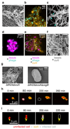
a, The curli fiber monomer CsgA was purified and shown by electron microscopy to polymerize in vitro (n=2). b, Curli fibers alone, visualized by immunostaining (orange), permit free diffusion of phage virions (cyan) into the curli mesh (n=3). c, Phages can bind to curli fibers that were polymerized in vitro before phage exposure, as indicated by the arrows (n=3). d, Fluorescent beads (magenta) were used as artificial replacements for bacterial cells. 1 μm diameter beads permit diffusion of phage virions throughout the interior of bead clusters. e, When beads are combined with curli polymers to produce minimal artificial biofilms, the bead clusters were protected from phage diffusion (n=4). f, Preformed curli fibers, when incubated with beads, localize between individual beads and wrap around bead clusters as shown by electron microscopy (n=3). g, Individual cells that are capable of curli production (but incapable of flagella and cellulose production) embed themselves in a dense mesh made out of self-produced curli fibers (n=3). h, Individual bacteria (red) that are surrounded by curli (yellow) are not infected by phages and can divide normally, whereas cells that are not surrounded by curli become infected (grey), and then lyse (white dashed line) (n=5). i, Bacteria that are not completely surrounded by curli are not protected from phage infection (n=5).

Comment in

  • Phaged and confused by biofilm matrix.
    Price JE, Chapman MR. Price JE, et al. Nat Microbiol. 2018 Jan;3(1):2-3. doi: 10.1038/s41564-017-0078-2. Nat Microbiol. 2018. PMID: 29255281 No abstract available.

References

    1. Flemming H-C, et al. Biofilms: an emergent form of bacterial life. Nat Rev Microbiol. 2016;14:563–75. - PubMed
    1. Persat A, et al. The mechanical world of bacteria. Cell. 2015;161:988–97. - PMC - PubMed
    1. Hobley L, Harkins C, MacPhee CE, Stanley-Wall NR. Giving structure to the biofilm matrix: an overview of individual strategies and emerging common themes. FEMS Microbiol Rev. 2015;39:649–69. - PMC - PubMed
    1. Berk V, et al. Molecular architecture and assembly principles of Vibrio cholerae biofilms. Science. 2012;337:236–9. - PMC - PubMed
    1. Drescher K, et al. Architectural transitions in Vibrio cholerae biofilms at single-cell resolution. Proc Natl Acad Sci U S A. 2016;113:E2066–72. - PMC - PubMed

MeSH terms

Substances

LinkOut - more resources