

Histopathological and genotypic characterization of metastatic colorectal carcinoma with PD-L1 (CD274)-expression: Possible roles of tumour micro environmental factors for CD274 expression
- PMID: 29085667
- PMCID: PMC5653930
- DOI: 10.1002/cjp2.81
Histopathological and genotypic characterization of metastatic colorectal carcinoma with PD-L1 (CD274)-expression: Possible roles of tumour micro environmental factors for CD274 expression
Abstract
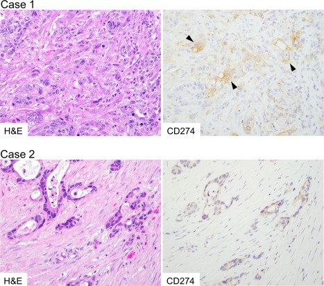
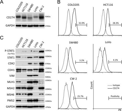
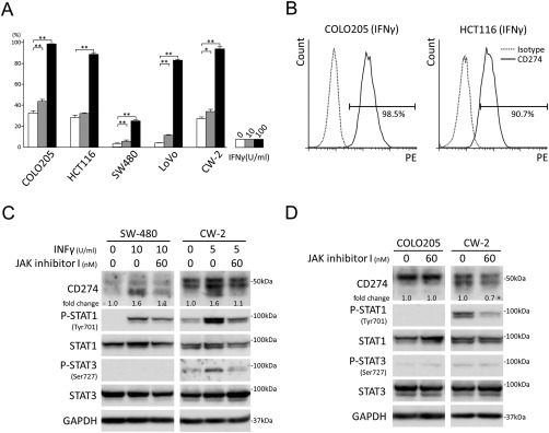
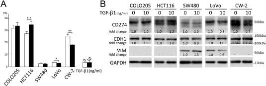
Aberrant PD-L1 (CD274) expression has been described in different types of tumour and linked to tumour aggressiveness and a poor prognosis. In primary colorectal carcinomas (CRCs), CD274 expression was reported to be associated with mismatch repair (MMR)-deficiency, BRAF mutation, and "stem-like" immunophenotype defined by down-regulation of homeobox protein CDX2 and membranous expression of activated leukocyte cell adhesion molecule (ALCAM). However, the immunophenotype and genotype of CD274-positive metastatic CRC have not been extensively analysed. In this study, 189 CRC metastases were evaluated immunohistochemically for CD274, MMR proteins, CDX2, and ALCAM expression. Immunostaining for CD4, CD8, and FOXP3 was also performed to characterize tumour-associated immune cells. In addition, 34 arbitrarily selected lesions were genotyped using Sanger- and next-generation sequencing. Univariate analyses showed no clear association between CD274 expression and clinicopathological parameters including MMR-deficiency or "stem-like" immunophenotype after adjustment for multiple testing. Comparison of the clinicopathological profiles of CD274-positive primary and metastatic tumours revealed in the latter younger age of occurrence (60.9 ± 13.3 versus 72.6 ± 13.1 years, p = 0.001), cytoplasm-dominant CD274 expression (p < 0.001), infrequent MMR-deficiency (p < 0.001), and common KRAS mutations (54%, p < 0.001). In five cultured colon cancer cell lines, CD274 was expressed and modulated after exogenous exposure to IFNγ and TGF-β1. Thus, CD274 regulation mechanisms might include tumour micro environmental factors. Based on significantly different characteristics in CD274-positive metastatic and primary CRCs, evaluation of metastases should also be considered when planning immune checkpoint inhibitor therapy.
Keywords: ALCAM (CD166); BRAF; CD274 (PD‐L1); CDX2; IFNγ; KRAS; TGF‐β1; immunohistochemistry; metastatic colorectal carcinoma.
Figures
References
-
- Nishimura H, Honjo T. PD‐1: an inhibitory immunoreceptor involved in peripheral tolerance. Trends Immunol 2001; 22 : 265–268. - PubMed
-
- Nishimura H, Nose M, Hiai H, et al Development of lupus‐like autoimmune diseases by disruption of the PD‐1 gene encoding an ITIM motif‐carrying immunoreceptor. Immunity 1999; 11 : 141–151. - PubMed
-
- Dong H, Zhu G, Tamada K, et al B7‐H1, a third member of the B7 family, co‐stimulates T‐cell proliferation and interleukin‐10 secretion. Nat Med 1999; 5 : 1365–1369. - PubMed
-
- Latchman Y, Wood CR, Chernova T, et al PD‐L2 is a second ligand for PD‐1 and inhibits T cell activation. Nat Immunol 2001; 2 : 261–268. - PubMed
LinkOut - more resources
Full Text Sources
Other Literature Sources
Research Materials
Miscellaneous
