

Syphilitic alopecia: uncommon trichoscopic findings
- PMID: 29085722
- PMCID: PMC5661157
- DOI: 10.5826/dpc.0703a12
Syphilitic alopecia: uncommon trichoscopic findings
Abstract
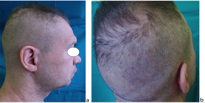
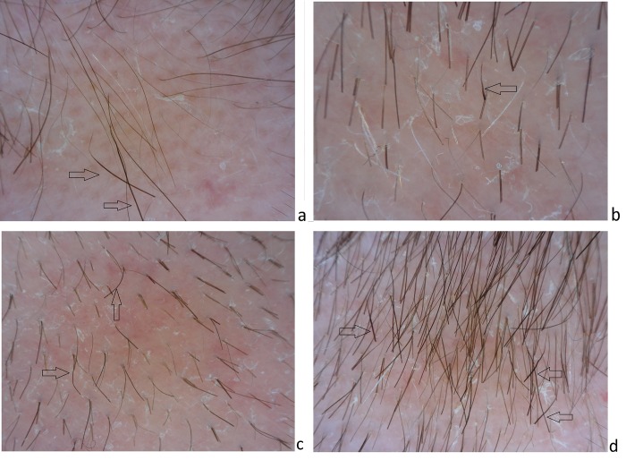
Syphilitic alopecia (SA) is considered an uncommon manifestation of secondary syphilis. SA can present in a diffuse form, resembling telogen effluvium, or in a moth-eaten form that mimics a variety of conditions (i.e., alopecia areata, trichotillomania, lichen planus pilaris or tinea capitis). When the two forms coexist, we observe a mixed pattern. Essential SA manifests without evidence of mucocutaneous syphilis manifestations and its diagnosis is often delayed. To date, trichoscopic description of SA forms are based on very few cases (i.e., five patients with moth-eaten SA and one with diffuse SA). This is the first report of a mixed pattern of essential SA: some new trichoscopic features-such as tapered bended hairs, erythematous background, diffuse scaling and perifollicular hyperkeratosis-are described in a 32-year-old man. In the absence of secondary syphilis manifestations, dermoscopy can be a useful tool that helps suspect and differentiate SA from its common mimickers.
Keywords: moth-eaten and diffuse syphilitic alopecia; secondary syphilis; trichoscopy.
Conflict of interest statement
Competing interests: The authors have no conflicts of interest to disclose.
Figures
References
-
- Vafaie J, Weinberg JM, Smith B, Mizuguchi RS. Alopecia in association with sexually transmitted disease: a review. Cutis. 2005;76:361–366. - PubMed
-
- Hernandez-Bel P, Unamuno B, Sanchez-Carazo JL, et al. Syphilitic alopecia: a report of 5 cases and a review of the literature. Actas Dermosifiliogr. 2013;104:512–517. - PubMed
-
- Bi MY, Cohen PR, Robinson FW, Gray JM. Alopecia syphilitica—report of a patient with secondary syphilis presenting as moth-eaten alopecia and a review of its common mimickers. Dermatol Online J. 2009;15:6. - PubMed
-
- Piraccini BM, Broccoli A, Starace M, et al. Hair and scalp manifestations in secondary syphilis: epidemiology, clinical features and trichoscopy. Dermatology. 2015;231(2):171–176. - PubMed
LinkOut - more resources
Full Text Sources
Other Literature Sources