

Impaired emotion processing in functional (psychogenic) tremor: A functional magnetic resonance imaging study
- PMID: 29085776
- PMCID: PMC5655406
- DOI: 10.1016/j.nicl.2017.10.020
Impaired emotion processing in functional (psychogenic) tremor: A functional magnetic resonance imaging study
Abstract
Background: Despite its high prevalence and associated disability, the neural correlates of emotion processing in patients with functional (psychogenic) tremor (FT), the most common functional movement disorder, remain poorly understood.
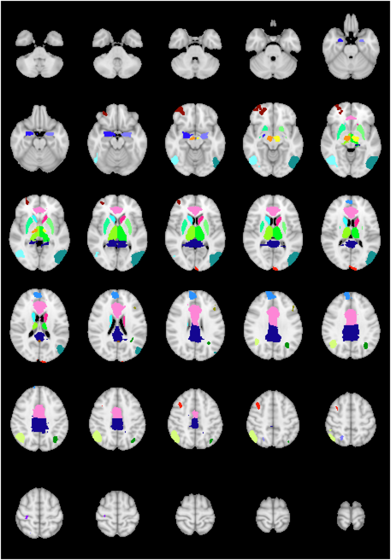
Methods: In this cross sectional functional magnetic resonance imaging (fMRI) study at 4T, 27 subjects with FT, 16 with essential tremor (ET), and 25 healthy controls (HCs) underwent a finger-tapping motor task, a basic-emotion task, and an intense-emotion task to probe motor and emotion circuitries. Anatomical and functional MRI data were processed with FSL (FMRIB Software Library) and AFNI (Analysis of Functional Neuroimages), followed by seed-to-seed connectivity analyses using anatomical regions defined from the Harvard-Oxford subcortical atlas; all analyses were corrected for multiple comparisons.
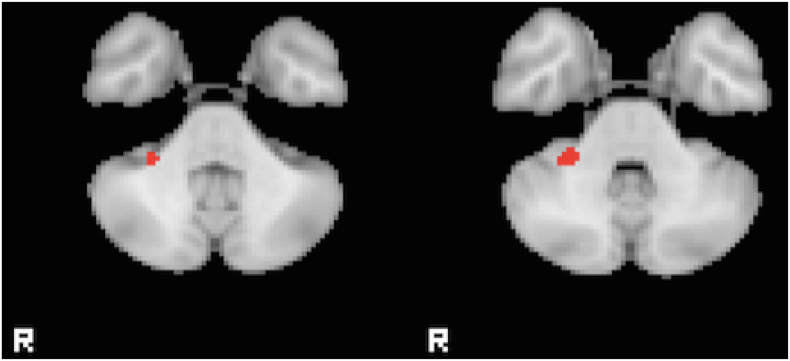
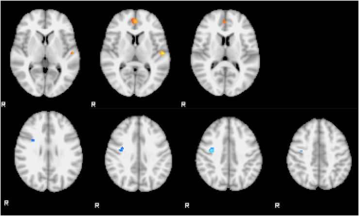
Results: After controlling for depression scores and correcting for multiple comparisons, the FT group showed increased activation in the right cerebellum compared to ET during the motor task; and increased activation in the paracingulate gyrus and left Heschl's gyrus compared with HC with decreased activation in the right precentral gyrus compared with ET during the basic-emotion task. No significant differences were found after adjusting for multiple comparisons during the intense-emotion task but increase in connectivity between the left amygdala and left middle frontal gyrus survived corrections in the FT subjects during this task, compared to HC.
Conclusions: In response to emotional stimuli, functional tremor is associated with alterations in activation and functional connectivity in networks involved in emotion processing and theory of mind. These findings may be relevant to the pathophysiology of functional movement disorders.
Keywords: AFNI, Analysis of Functional Neuroimages; CPT-END, continuous performance task with emotional and neutral distracters; Conversion disorder; EPI, echo-planar imaging; Emotion processing; FSL, FMRIB Software Library; FT, functional tremor; Functional movement disorders; Functional tremor; HAM-A, Hamilton Anxiety Rating Scale; HAM-D, Hamilton Depression Rating Scale; MDEFT, modified equilibrium Fourier transform; MINI, Mini International Neuropsychiatric Interview; Psychogenic tremor; fMRI; fMRI, functional magnetic resonance imaging.
Figures
References
-
- Allendorfer J.B., Szaflarski J.P. Contributions of fMRI towards our understanding of the response to psychosocial stress in epilepsy and psychogenic nonepileptic seizures. Epilepsy Behav. 2014;35C:19–25. - PubMed
-
- Andersson M.J.a.S.S. 2007. Non-linear Registration, aka Spatial Normalisation.
-
- Bar-On R., Tranel D., Denburg N.L., Bechara A. Exploring the neurological substrate of emotional and social intelligence. Brain. 2003;126:1790–1800. - PubMed
-
- Bartsch K., Estes D. Why we assume it's all good: the role of theory of mind in early inherent feature inferences. Behav. Brain Sci. 2014;37:482. - PubMed
MeSH terms
Grants and funding
LinkOut - more resources
Full Text Sources
Other Literature Sources
Medical
