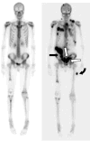
Unusual abdominal metastases in osteosarcoma
- PMID: 29085778
- PMCID: PMC5659360
- DOI: 10.1016/j.epsc.2017.09.022
Unusual abdominal metastases in osteosarcoma
Abstract
Intraabdominal metastases in the setting of osteosarcoma are very rare. We describe a case of a 17-year-old boy with high-grade right distal femur osteosarcoma who two years after diagnosis developed extensive intra abdominal metastases involving the omentum, peritoneum, bowel serosa, psoas muscles and abdominal soft tissue. Awareness of and surveillance for unusual patterns of metastasis may allow for earlier detection, intervention, and palliative care decision-making, which may affect survival and quality of life. This report underlines the need for prospective studies evaluating surveillance guidelines for patients after medical and surgical management of osteosarcoma, especially in cases complicated by pulmonary metastases.
Keywords: Pediatric osteosarcoma; extra-pulmonary metastases; extra-pulmonary recurrence; surveillance imaging.
Conflict of interest statement
Conflict of Interest Statement: The authors declare that there are no known conflicts of interest associated with this publication and there has been no significant financial support for this work that could have influenced its outcome.
Figures


References
-
- Ottaviani G, Jaffe N. The etiology of osteosarcoma. In: Jaffe N, Bruland OS, Bielack S, editors. Pediatric and Adolescent Osteosarcoma. New York: Springer; 2009. pp. 15–32. - PubMed
-
- Gurney JG, Swensen AR, Bulterys M. Malignant bone tumors. In: Ries LA, Smith MA, Gurney JG, et al., editors. Cancer Incidence and Survival Among Children and Adolescents: United States SEER Program 1975–1995. Bethesda, MD: National Cancer Institute, SEER Program; 1999. pp. 99–110. NIH Pub. No. 99-4649.
-
- Bleyer A, O’Leary M, Barr R, Ries LAG, editors. Cancer Epidemiology in Older Adolescents and Young Adults 15 to 29 Years of Age, Including SEER Incidence and Survival: 1975–2000. Bethesda, MD: National Cancer Institute; 2006. NIH Pub. No. 06-5767.
-
- Mialou V, Philip T, Kalifa C, Perol D, Gentet JC, Marec-Berard P, Pacquement H, Chastagner P, Defaschelles AS, Hartmann O. Metastatic osteosarcoma at diagnosis: prognostic factors and long-term outcome--the French pediatric experience. Cancer. 2005 Sep 1;104(5):1100–1109. doi: 10.1002/cncr.21263. - DOI - PubMed
Grants and funding
LinkOut - more resources
Full Text Sources
Other Literature Sources
