

Adrenal ganglioneuroma: What you need to know
- PMID: 29085827
- PMCID: PMC5648998
- DOI: 10.12998/wjcc.v5.i10.373
Adrenal ganglioneuroma: What you need to know
Abstract
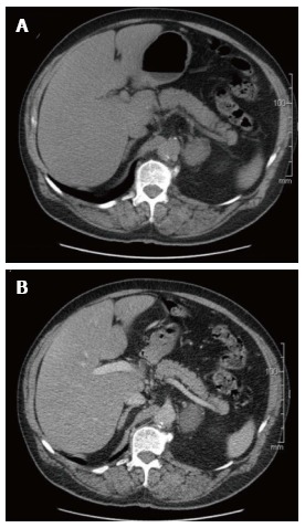
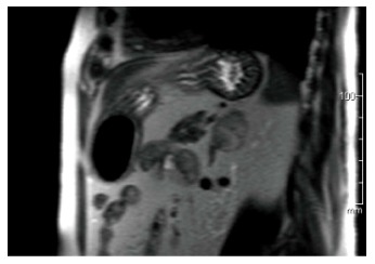
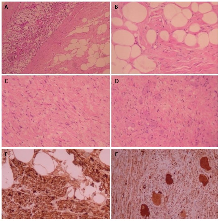
Adrenal ganglioneuromas (GNs) constitute rare, differentiated tumors which originate from neural crest cells. GNs are usually hormonally silent and tend to be discovered incidentally on imaging tests. Adrenalectomy is the gold standard for the treatment of primary adrenal GNs. Nevertheless, preoperative differential diagnosis of GNs remains extremely challenging, and thus histopathological examination is required in order to confirm the diagnosis of GN. Overall, prognosis after surgical resection seems to be excellent, without any recurrences or need for adjuvant therapy.
Keywords: Adrenalectomy; Ganglioneuroma; Neural crest; Neurogenic tumors.
Conflict of interest statement
Conflict-of-interest statement: The authors declare no potential conflicts of interest with respect to the research, authorship, and/or publication of this article.
Figures
References
-
- Joshi VV. Peripheral neuroblastic tumors: pathologic classification based on recommendations of international neuroblastoma pathology committee (Modification of shimada classification) Pediatr Dev Pathol. 2000;3:184–199. - PubMed
-
- Rha SE, Byun JY, Jung SE, Chun HJ, Lee HG, Lee JM. Neurogenic tumors in the abdomen: tumor types and imaging characteristics. Radiographics. 2003;23:29–43. - PubMed
-
- Geoerger B, Hero B, Harms D, Grebe J, Scheidhauer K, Berthold F. Metabolic activity and clinical features of primary ganglioneuromas. Cancer. 2001;91:1905–1913. - PubMed
-
- Qing Y, Bin X, Jian W, Li G, Linhui W, Bing L, Huiqing W, Yinghao S. Adrenal ganglioneuromas: a 10-year experience in a Chinese population. Surgery. 2010;147:854–860. - PubMed
Publication types
LinkOut - more resources
Full Text Sources
Other Literature Sources
