. 2018 Feb;36(2):265-277.
doi: 10.1002/stem.2732. Epub 2017 Nov 13.

Passive Stretch Induces Structural and Functional Maturation of Engineered Heart Muscle as Predicted by Computational Modeling

Affiliations

Passive Stretch Induces Structural and Functional Maturation of Engineered Heart Muscle as Predicted by Computational Modeling

Oscar J Abilez et al. Stem Cells. 2018 Feb.

Abstract

The ability to differentiate human pluripotent stem cells (hPSCs) into cardiomyocytes (CMs) makes them an attractive source for repairing injured myocardium, disease modeling, and drug testing. Although current differentiation protocols yield hPSC-CMs to >90% efficiency, hPSC-CMs exhibit immature characteristics. With the goal of overcoming this limitation, we tested the effects of varying passive stretch on engineered heart muscle (EHM) structural and functional maturation, guided by computational modeling. Human embryonic stem cells (hESCs, H7 line) or human induced pluripotent stem cells (IMR-90 line) were differentiated to hPSC-derived cardiomyocytes (hPSC-CMs) in vitro using a small molecule based protocol. hPSC-CMs were characterized by troponin+ flow cytometry as well as electrophysiological measurements. Afterwards, 1.2 × 10⁶ hPSC-CMs were mixed with 0.4 × 10⁶ human fibroblasts (IMR-90 line) (3:1 ratio) and type-I collagen. The blend was cast into custom-made 12-mm long polydimethylsiloxane reservoirs to vary nominal passive stretch of EHMs to 5, 7, or 9 mm. EHM characteristics were monitored for up to 50 days, with EHMs having a passive stretch of 7 mm giving the most consistent formation. Based on our initial macroscopic observations of EHM formation, we created a computational model that predicts the stress distribution throughout EHMs, which is a function of cellular composition, cellular ratio, and geometry. Based on this predictive modeling, we show cell alignment by immunohistochemistry and coordinated calcium waves by calcium imaging. Furthermore, coordinated calcium waves and mechanical contractions were apparent throughout entire EHMs. The stiffness and active forces of hPSC-derived EHMs are comparable with rat neonatal cardiomyocyte-derived EHMs. Three-dimensional EHMs display increased expression of mature cardiomyocyte genes including sarcomeric protein troponin-T, calcium and potassium ion channels, β-adrenergic receptors, and t-tubule protein caveolin-3. Passive stretch affects the structural and functional maturation of EHMs. Based on our predictive computational modeling, we show how to optimize cell alignment and calcium dynamics within EHMs. These findings provide a basis for the rational design of EHMs, which enables future scale-up productions for clinical use in cardiovascular tissue engineering. Stem Cells 2018;36:265-277.

Keywords: Bioengineering; Calcium handling; Cardiac; Cardiomyocyte; Computational modeling; Engineered heart muscle; Heart; Pluripotent stem cells; Tissue engineering; Tissue regeneration.


Conflict of interest statement

Disclosure of Potential Conflicts of Interest

J.R. is an employee of Genentech. Evangeline Tzatzalos discloses an advisory role with Wilson Sonsini Goodrich and is an employee of Rosati. D.B. is a patent holder for RegenCor (collagen patch for cardiac regeneration). All other authors indicated no potential conflicts of interest.

Figures

Figure 1
Figure 1
Computational modeling reveals spatial distribution of maximum principal stress in engineered heart muscle (EHM) for varying passive stretch and EHM compaction. (A): EHMs show low stresses in the center and the unpopulated areas next to the post holes, with the initial assumption that EHMs detach from the inner surface of the end posts during formation. V-necking is apparent at the site of the posts. (B): EHMs show higher stress in the center and increased thinning for increased EHM passive stretch, with the initial assumption that EHMs stay attached to the inner surface of the end posts during formation. Percentages along the top are volume compaction; baseline (no stretch), 5, 7, and 9 mm are the distances between the posts. Dashed rectangles highlight optimal geometries seen in experiments. Color code from 0.00 to 1.00 shows normalized stresses compared with baseline.
Figure 2
Figure 2
Generation and formation of engineered heart muscles (EHMs) of varying passive stretch. (A): Human pluripotent stem cell-derived cardiomyocytes and human fibroblasts (IMR-90 line) were mixed with type-I collagen to create EHMs. (B): The cell/collagen mixture was seeded into polydimethylsiloxane (PDMS) molds which contain five 100 μl reservoirs and fit within the wells of a six-well dish. (C): EHM were cast in reservoirs containing two PDMS posts spaced at 5, 7, and 9 mm or containing only one post (control with no tension). EHMs were allowed to condense and were analyzed at various time points within days 1–50 by quantitative polymerase chain reaction, immunohistochemistry, calcium imaging, and force testing. Abbreviations: CM, cardiomyocytes; EHM, engineered heart muscle; hPSC, human pluripotent stem cell; IHC, immunohistochemistry; PDMS, polydimethylsiloxane; qPCR, quantitative polymerase chain reaction.
Figure 3
Figure 3
Engineered heart muscle (EHM) formation depends on passive stretch. (A): EHMs were observed daily for wall detachment, post detachment, opacity, compaction, and V-neck formation. (B): Human pluripotent stem cell-derived cardiomyocytes were mixed with fibroblasts (IMR-90 line) and type-I collagen and seeded into custom-designed PDMS molds. EHM formation was monitored over time (days 0–5). Inter-post distance of a representative EHM is 7 mm. A single post (conferring no tension) served as a control. White scale bar = 7 mm. Daily observations of EHM formation according to predetermined criteria by percentage EHMs, including (C) detachment from the reservoir walls, (D) detachment from posts, (E) opacity, (F) extent of compaction, and (G, H) formation of a tension-induced V-neck at EHM-anchoring end posts. White scale bar = 1 mm (H). Abbreviations: EHM, engineered heart muscle; hPSC, human pluripotent stem cell; PDMS, polydimethylsiloxane.
Figure 4
Figure 4
Low magnification immunohistochemistry shows cardiovascular cell types are present in engineered heart muscles (EHMs) and distribution depends on passive stretch. (A–D): At the EHM middle, cardiomyocytes labeled with α-actinin (red) and troponin-T (green) are present throughout the EHM width, with Troponin-T expressed most highly at the EHM outer edges. (E–G): At the EHM ends, cardiomyocytes labeled with α-actinin (red) and troponin-T (green) are also present throughout the EHM width, with troponin-T expressed most highly at the EHM outer edges. (H–K): At the EHM middle, fibroblasts labeled with FSP-1 (red) and endothelial cells labeled with CD31 (green) are homogeneously present throughout the EHM width. (L–N): At the EHM ends, fibroblasts labeled with FSP-1 (red) and endothelial cells labeled with CD31 (green) are also homogeneously present throughout the EHM width. (C, F, J, M) The 7 mm passive stretch shows the most organized morphology (dashed green rectangles). In all panels, 4′,6-diamidino-2-phenylindole labels cellular nuclei. White scale bar = 100 μm (A, E, L, N). Objective magnification is ×10. Abbreviations: DAPI, 4′,6-diamidino-2-phe-nylindole; EHM, engineered heart muscle; FSP-1, fibroblast specific protein-1.
Figure 5
Figure 5
High-magnification immunohistochemistry shows cardiovascular cell types are present in engineered heart muscles (EHMs) with various sarcomeric organization at different passive lengths. (A–D): At the EHM middle and outer edges, cardiomyocytes labeled with α-actinin (red) and troponin-T (green) are present, with sarcomeric banding patterns visible at all lengths as shown in insets. (E–H): At the EHM middle and outer edges, fibroblasts labeled with FSP-1 (red) and endothelial cells labeled with CD31 (green) are present. (I): Representative images of day 30 EHMs show various degrees of cardiomyocyte sarcomeric alignment at control, 5, 7, and 9 mm passive stretch as demonstrated by α-actinin (green) and troponin-T (red) immunostaining. White double-ended arrows indicate the primary direction of stretch. (J): High-magnification views of the area insets indicated by the white dashed rectangles in (I). White double-ended arrows indicate the primary direction of stretch while yellow double-ended dashed arrows indicate the general direction of sarcomeric alignment. EHMs under 7 mm stretch demonstrated sarcomeres most aligned with the primary direction of stretch as compared with control, 5, and 9 mm groups. In all panels, 4′,6-diamidino-2-phenylindole labels cellular nuclei. White scale bar = 20 μm (A, E). Objective magnification is ×63 in (A–H) and ×40 in (I). Abbreviations: DAPI, 4′,6-diamidino-2-phenylindole; EHM, engineered heart muscle; FSP-1, fibroblast specific protein-1.
Figure 6
Figure 6
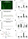
Calcium dynamics correlates with engineered heart muscle (EHM) passive stretch. (A): Representative image of Fluo-4 staining to detect calcium transients. Objective magnification is ×10. (B, C): Relative Fluo-4 intensities over time with and without electrical stimulation at 1 Hz. (D): Calcium imaging waveform analytical parameters. Analytical outputs for spontaneous and electrically paced EHMs at 5, 7, and 9 mm interpost distances: (E) amplitude, (F) time-to-peak, (G) beating rate, and (H) TD50. Each colored waveform in (B) is a representative waveform of the spontaneous, non-paced calcium dynamics of EHMs (n = 12) generated under tension at 7 mm stretch. The mean beating rates of all stretch conditions for both spontaneous and paced groups are shown in (G). Mean ± SEM.*, p < .05 (Student’s t test); #, p < .05 (one-way analysis of variance with Tukey’s post hoc testing).
Figure 7
Figure 7
Engineered heart muscle (EHM) gene expression indicates increased maturation of cardiomyocytes. (A): EHMs (7 mm inter-post distance) were formed with day 22 cardiomyocytes and were matured until days 25, 30, and 50. (B): Quantitative polymerase chain reaction analysis was performed using primers for β1-adrenergic receptor (ADRB1), calveolin-3 (CAV3), potassium ion channel (KCNJ2), β2-adrenergic receptor (ADRB2), troponin-T (TNNT2), and calcium ion channel (CACNA1C). Gene expression in EHM+ samples were normalized by the ratio of cardiomyocytes to fibroblast (3:1). Compared with day 50 cardiomyocytes, day 50 expression of ADRB1, CAV3, TNNT2, and CACNA1C of EHMs with fibroblasts (EHM1) was all statistically significantly increased. Mean ± SEM. *, p < .05 versus day 50 monolayer cardiomyocytes (one-way analysis of variance with Tukey’s post hoc testing). Abbreviations: CM, cardiomyocytes; EHM, engineered heart muscle; iPSC, induced pluripotent stem cell.

References

    1. Lloyd-Jones D, Adams RJ, Brown TM, et al. Heart disease and stroke statistics– 2010 update: A report from the American Heart Association. Circulation. 2010;121:e46–e215. - PubMed
    1. Laflamme MA, Murry CE. Heart regeneration. Nature. 2011;473:326–335. - PMC - PubMed
    1. Velagaleti RS, Pencina MJ, Murabito JM, et al. Long-term trends in the incidence of heart failure after myocardial infarction. Circulation. 2008;118:2057–2062. - PMC - PubMed
    1. Healy KE, McDevitt TC, Murphy WL, et al. Engineering the emergence of stem cell therapeutics. Sci Transl Med. 2013;5:207ed217. - PubMed
    1. Ye L, Zimmermann WH, Garry DJ, et al. Patching the heart: Cardiac repair from within and outside. Circ Res. 2013;113:922–932. - PMC - PubMed

Publication types