. 2017 Oct;22(10):1-10.
doi: 10.1117/1.JBO.22.10.106017.

Combining deep learning and coherent anti-Stokes Raman scattering imaging for automated differential diagnosis of lung cancer

Affiliations

Combining deep learning and coherent anti-Stokes Raman scattering imaging for automated differential diagnosis of lung cancer

Sheng Weng et al. J Biomed Opt. 2017 Oct.

Abstract

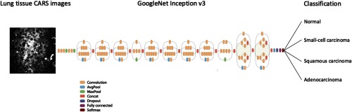
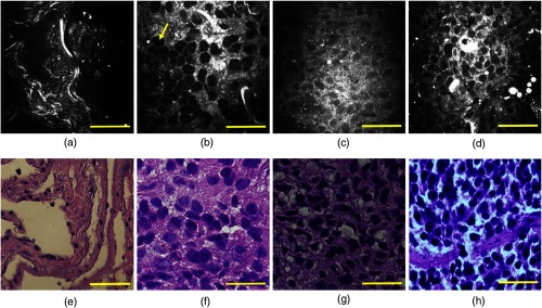
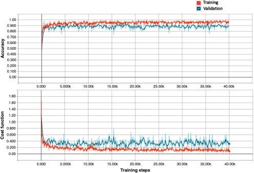
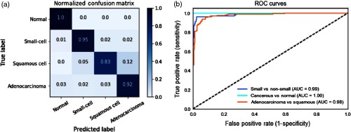
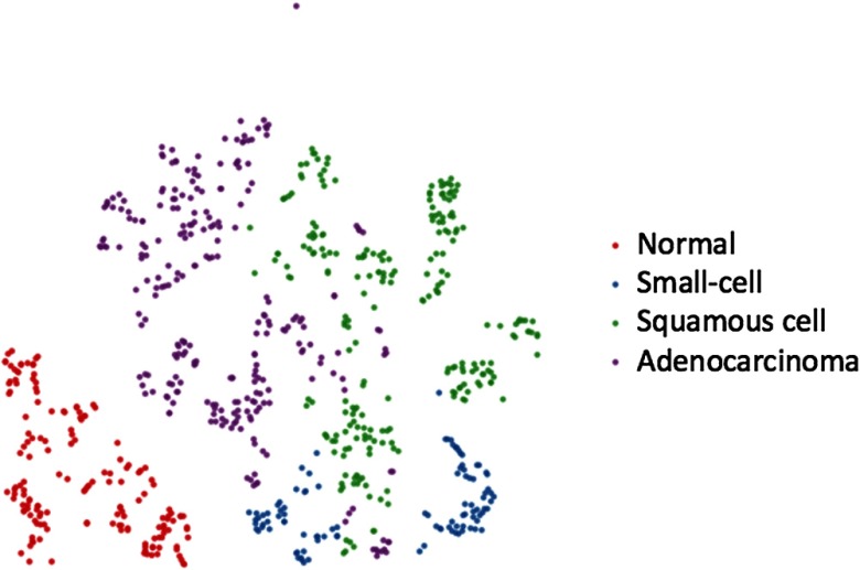
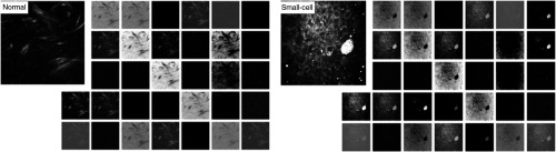
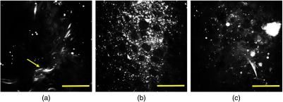
Lung cancer is the most prevalent type of cancer and the leading cause of cancer-related deaths worldwide. Coherent anti-Stokes Raman scattering (CARS) is capable of providing cellular-level images and resolving pathologically related features on human lung tissues. However, conventional means of analyzing CARS images requires extensive image processing, feature engineering, and human intervention. This study demonstrates the feasibility of applying a deep learning algorithm to automatically differentiate normal and cancerous lung tissue images acquired by CARS. We leverage the features learned by pretrained deep neural networks and retrain the model using CARS images as the input. We achieve 89.2% accuracy in classifying normal, small-cell carcinoma, adenocarcinoma, and squamous cell carcinoma lung images. This computational method is a step toward on-the-spot diagnosis of lung cancer and can be further strengthened by the efforts aimed at miniaturizing the CARS technique for fiber-based microendoscopic imaging.

Keywords: artificial intelligence; classification; deep learning; lung cancer; medical imaging; nonlinear microscopy.


Figures

Fig. 1
Fig. 1
Transfer learning layout. Data flow is from left to right: a CARS image of human lung tissue is fed into GoogleNet Inception v3 CNN pretrained on the ImageNet data and fine-tuned on our own CARS images comprising four classes: normal, small-cell carcinoma, squamous carcinoma, and adenocarcinoma. The model outputs the probability distribution over the four classes and we label the CARS image with the largest probability class. GoogleNet Inception v3 CNN architecture reprinted from Ref. .
Fig. 2
Fig. 2
Representative CARS images (upper panels) and corresponding H&E-stained images (lower panels) of human lung tissues: (a) and (e) normal lung, (b) and (f) adenocarcinoma, (c) and (g) squamous cell carcinoma, and (d) and (h) small-cell carcinoma. Scale bars: 50  μm.
Fig. 3
Fig. 3
Learning curves of training and validation for one cross-validation round visualized by TensorBoard. The short-term noise is due to the stochastic gradient descent nature of the algorithm. The curves are smoothed for better visualization.
Fig. 4
Fig. 4
Deep CNN model performance on the holdout test set. (a) Normalized confusion matrix. Each row represents the instances in a ground-truth class and the value in each column represents what percentage of the images is predicted to a certain class. (b) ROC curves for three conditions: separating cancerous from normal lung images (light blue); separating small-cell carcinoma from nonsmall cell carcinoma lung images (dark blue); separating adenocarcinoma and squamous carcinoma lung images (orange). AUC scores are given in the legend.
Fig. 5
Fig. 5
t-SNE visualization of the last hidden layer representations in the deep CNN of the holdout test set (758-CARS images). Colored point clouds represent four different human lung tissue classes clustered by the algorithm. Red points are normal lung images, blue points are small-cell carcinoma images, green points are squamous cell carcinoma images, and purple points are adenocarcinoma images.
Fig. 6
Fig. 6
Visualization of the first convolutional layer in GoogleNet Inception v3 model for a normal and a small-cell lung carcinoma CARS images. The first convolutional layer contains 32 kernels with a size of 149×149  pixels.
Fig. 7
Fig. 7
Representative CARS images of human lung tissues misclassified by the algorithm. (a) Adenocarcinoma is labeled as normal lung, (b) squamous cell carcinoma is labeled as adenocarcinoma, and (c) small-cell carcinoma is labeled as squamous cell carcinoma. Scale bars: 50  μm.

References

    1. Siegel R. L., Miller K. D., Jemal A., “Cancer statistics, 2017,” CA. Cancer J. Clin. 67, 7–30 (2017).CAMCAM10.3322/caac.21387 - DOI - PubMed
    1. Youlden D. R., Cramb S. M., Baade P. D., “The international epidemiology of lung cancer: geographical distribution and secular trends,” J. Thorac. Oncol. 3, 819–831 (2008).10.1097/JTO.0b013e31818020eb - DOI - PubMed
    1. McWilliams A., et al. , “Innovative molecular and imaging approaches for the detection of lung cancer and its precursor lesions,” Oncogene 21, 6949–6959 (2002).ONCNES10.1038/sj.onc.1205831 - DOI - PubMed
    1. Cagle P. T., et al. , “Revolution in lung cancer: new challenges for the surgical pathologist,” Arch. Pathol. Lab. Med. 135, 110–116 (2011).APLMAS10.1043/2010-0567-RA.1 - DOI - PubMed
    1. Henschke C. I., et al. , “Early lung cancer action project: overall design and findings from baseline screening,” Lancet 354, 99–105 (1999).LANCAO10.1016/S0140-6736(99)06093-6 - DOI - PubMed

MeSH terms