. 2017 Nov 14;114(46):E9913-E9922.
doi: 10.1073/pnas.1707779114. Epub 2017 Oct 30.

Relevance of iPSC-derived human PGC-like cells at the surface of embryoid bodies to prechemotaxis migrating PGCs

Affiliations

Relevance of iPSC-derived human PGC-like cells at the surface of embryoid bodies to prechemotaxis migrating PGCs

Shino Mitsunaga et al. Proc Natl Acad Sci U S A. .

Abstract

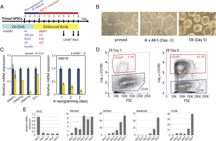
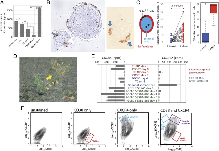
Pluripotent stem cell-derived human primordial germ cell-like cells (hPGCLCs) provide important opportunities to study primordial germ cells (PGCs). We robustly produced CD38+ hPGCLCs [∼43% of FACS-sorted embryoid body (EB) cells] from primed-state induced pluripotent stem cells (iPSCs) after a 72-hour transient incubation in the four chemical inhibitors (4i)-naïve reprogramming medium and showed transcriptional consistency of our hPGCLCs with hPGCLCs generated in previous studies using various and distinct protocols. Both CD38+ hPGCLCs and CD38- EB cells significantly expressed PRDM1 and TFAP2C, although PRDM1 mRNA in CD38- cells lacked the 3'-UTR harboring miRNA binding sites regulating mRNA stability. Genes up-regulated in hPGCLCs were enriched for cell migration genes, and their promoters were enriched for the binding motifs of TFAP2 (which was identified in promoters of T, NANOS3, and SOX17) and the RREB-1 cell adhesion regulator. In EBs, hPGCLCs were identified exclusively in the outermost surface monolayer as dispersed cells or cell aggregates with strong and specific expression of POU5F1/OCT4 protein. Time-lapse live cell imaging revealed active migration of hPGCLCs on Matrigel. Whereas all hPGCLCs strongly expressed the CXCR4 chemotaxis receptor, its ligand CXCL12/SDF1 was not significantly expressed in the whole EBs. Exposure of hPGCLCs to CXCL12/SDF1 induced cell migration genes and antiapoptosis genes. Thus, our study shows that transcriptionally consistent hPGCLCs can be readily produced from hiPSCs after transition of their pluripotency from the primed state using various methods and that hPGCLCs resemble the early-stage PGCs randomly migrating in the midline region of human embryos before initiation of the CXCL12/SDF1-guided chemotaxis.

Keywords: PRDM1; TFAP2C; embryoid body; induced pluripotent stem cells; primordial germ cells.


Conflict of interest statement

The authors declare no conflict of interest.

Figures

Fig. 1.
Fig. 1.
Production of hPGCLCs from primed pluripotency hiPSCs via 72-h reprogramming toward naïve pluripotency. (A) Diagram of the 11-d schedule of hPGCLC production from primed pluripotency hiPSCs. (B) Phase contrast images of hiPSCs before and after reprograming toward naïve pluripotency in the 4i medium and day 5 EBs. (C) mRNA expression of TET1 and DNA methyltransferase genes in the primed and 4i reprogrammed hiPSCs. TaqMan real-time qPCR measurements (n = 3, mean ± SD). (D) FACS enrichment of CD38+ and CD38 EB cells from days 5 and 8 EBs. FSC, forward scatter. (E) mRNA expression of germline markers in EBs. TaqMan real-time qPCR measurements (n = 6, mean ± SD).
Fig. 2.
Fig. 2.
Transcriptomal profiles of CD38+ hPGCLCs and precursors. (A and B) Unsupervised hierarchical clustering of CD38+ hPGCLCs and their precursors based on transcriptomes (A) or 24 marker genes (B). Details of the three gene clusters 1–3 indicated in A are shown in Fig. S1 C1–C3, respectively. RNA-seq data were obtained from three independently performed experiments. Gray color indicates “zero value” data, reflecting the absence of expression. (C) Transcriptomal PCA. Arrows indicate the path of hiPSC differentiation to CD38+ hPGCLCs and CD38 EB cells. (D) RNA-seq tracks at the 3′-UTR of PRDM1 mRNA. Positions of exons 6–8, 3′-UTR, and miRNA binding sites are indicated. (E) GO analysis of DEGs between CD38+ hPGCLCs and CD38 EB cells.
Fig. 3.
Fig. 3.
Marker gene expression profiles of human germ cells, hPGCLCs, and precursors across various protocols. (A) Unsupervised hierarchical clustering of human embryonic and in vitro germline cells generated in four independent laboratories using different protocols. Clustering was performed based on 46 marker genes of pluripotency (blue), PGC (red), and AGCs (green). Color-coded cell types shown at the top of the heat map are indicated in B. Groups of genes expressed in the (1) pluripotent precursor cells, (2) nongermline EB cells (CD38), (3) hPGCLCs, and (4) AGCs are shown with yellow rectangles. Gray color indicates zero value data, reflecting the absence of expression. GS, gonadal somatic cells; TC, TCam-2 human seminoma cells. (B) Color identifications of transcriptomal data. Data from this study are indicated by red caps in B and circles in C. Data reported by Sasaki et al. (16), Irie et al. (15), and Gkountela et al. (23) are indicated in green, blue, and orange, respectively. (C) PCA of human embryonic and in vitro germline cells with data shown in A. PC, principal component.
Fig. 4.
Fig. 4.
TF binding motifs enriched in the regulatory sequences of 537 DEGs expressed more strongly in CD38+ hPGCLCs than in CD38 EB cells. (A) List of significantly enriched TFs (P < 0.01). The P value was defined by the F-match algorithm and calculated by the TRANSFAC server. (B) Locations of the TFAP2 binding motifs in promoters of NANOS3, SOX17, and T/BRACHYURY. Matches to the core motif (GCCNNNGGC) are shown in red, and matches to the additional contributing sequences are shown in orange.
Fig. 5.
Fig. 5.
Localization of CD38+ hPGCLCs at the outermost surface layer of EBs. (A) Expression of POU5F1 mRNA transcripts: RNA-seq (n = 3, mean ± SEM). Statistical significance (t test) is indicated by asterisks. *P < 0.05; **P < 0.01; ***P < 0.001. (B) Localization of POU5F1+ hPGCLCs at the outermost surface layer of day 8 EBs. hPGCLCs were observed in formaldehyde-fixed, paraffin-embedded EBs by immunohistochemistry. (Left) hPGCLCs distributed at the outermost surface monolayer of EBs (POU5F1 protein is stained brown with hematoxylin counterstaining). (Right) hPGCLCs are found as aggregates at EB surface (no counterstaining). Arrows indicate aggregated cells strongly expressing POU5F1. (C) Statistical evaluation of surface enrichment of POU5F1++ (strongly positive) hPGCLCs. (Left) Definition of the “surface layer,” which is the outermost surface monolayer of EBs, and the “internal area,” which is the remaining region. (Center) Number of POU5F1++ cells in the internal or surface regions of each EB (n = 23; P value was calculated by paired t test). (Right) Box plot representation of percentage localization of POU5F1++ cells in the internal or surface regions of EBs. (D) Time-lapse live imaging of a CD38+ hPGCLC showing active cell migration. A live EB was immobilized on Matrigel overnight, and an hPGCLC that emerged from the EB mass was stained with a fluorescence dye-conjugated anti-CD38 antibody. Fluorescence images were taken before and after a 60-min time lapse and digitally converted to green and orange pseudocolors, respectively. The CD38+ hPGCLC marked as a showed a well-developed cellular protrusion (arrowheads) and moved to the location marked as b within 60 min. (E) Expression of the CXCR4 chemotaxis receptor and its ligand CXCL12 in human germline cells and gonadal somatic cells. RNA-seq data generated in four independent laboratories were conormalized to obtain relative expression profiles (of CXCR4 and CXCL12 mRNA across cell types. cpm, Counts per million. (F) FACS analysis of total day 5 EB cells for cell surface expression of CD38 and CXCR4. The double-stained FACS chart indicates gates for single- and double-positive cells.
Fig. 6.
Fig. 6.
Effects of CXCL12 on mRNA expression in hPGCLCs. (A) Expression of mRNA transcripts in CD38+ hPGCLCs or CD38 EB cells at day 8 EBs. EBs were cultured in the standard condition until day 5 and then incubated for an additional 3 d in the presence or absence of Rho Kinase Inhibitor (ROCKi) and/or CXCL12 in the PGCLC production medium. Amounts of mRNA expression are shown as counts per million (cpm) values of normalized reads of three repeated RNA-seq experiments. (B and C) GO analysis of DEGs in hPGCLCs produced in the ROCKi-deficient medium in the presence or absence of CXCL12. Three RNA-seq experiments determined 1,190 up-regulated (B) and 735 down-regulated (C) DEGs. Statistically significant GO terms (FDR < 5%) are shown. (D) RNA-seq traces of CXCR4, PTGER3, BCL2, and CASP8 mRNA expression. The height of peaks represents relative mRNA expression of a gene in the six cell culture conditions and was normalized for each gene. ZRAMB2 locates adjacent to PTGER3 and is shown as control. KDSR and VPS4B are controls located adjacent to BCL2. ACTB expression shows quantitative reproducible RNA-seq experiments across the six different cell culture conditions. Note that PTGER3 and BCL2 traces are shown only at representative exons.

References

    1. Weinberger L, Ayyash M, Novershtern N, Hanna JH. Dynamic stem cell states: Naive to primed pluripotency in rodents and humans. Nat Rev Mol Cell Biol. 2016;17:155–169. - PubMed
    1. Gafni O, et al. Derivation of novel human ground state naive pluripotent stem cells. Nature. 2013;504:282–286. - PubMed
    1. Theunissen TW, et al. Systematic identification of culture conditions for induction and maintenance of naive human pluripotency. Cell Stem Cell. 2014;15:471–487. - PMC - PubMed
    1. Choi J, et al. Prolonged Mek1/2 suppression impairs the developmental potential of embryonic stem cells. Nature. 2017;548:219–223. - PMC - PubMed
    1. Magnúsdóttir E, Surani MA. How to make a primordial germ cell. Development. 2014;141:245–252. - PubMed

Publication types

MeSH terms

Substances