. 2017 Nov 14;114(46):12196-12201.
doi: 10.1073/pnas.1708744114. Epub 2017 Oct 30.

DsbA-L prevents obesity-induced inflammation and insulin resistance by suppressing the mtDNA release-activated cGAS-cGAMP-STING pathway

Affiliations

DsbA-L prevents obesity-induced inflammation and insulin resistance by suppressing the mtDNA release-activated cGAS-cGAMP-STING pathway

Juli Bai et al. Proc Natl Acad Sci U S A. .

Abstract

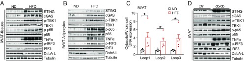
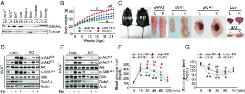
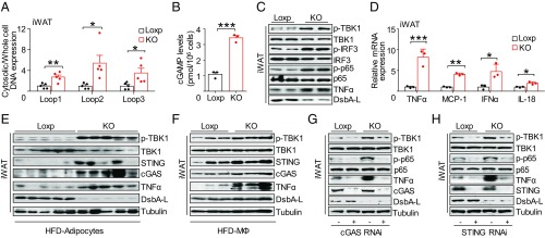
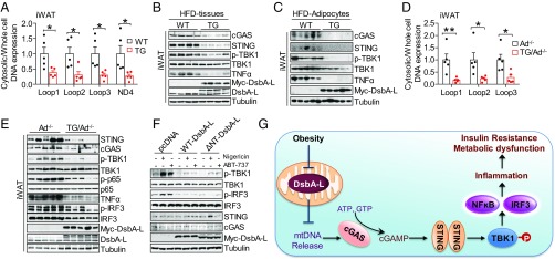
Chronic inflammation in adipose tissue plays a key role in obesity-induced insulin resistance. However, the mechanisms underlying obesity-induced inflammation remain elusive. Here we show that obesity promotes mtDNA release into the cytosol, where it triggers inflammatory responses by activating the DNA-sensing cGAS-cGAMP-STING pathway. Fat-specific knockout of disulfide-bond A oxidoreductase-like protein (DsbA-L), a chaperone-like protein originally identified in the mitochondrial matrix, impaired mitochondrial function and promoted mtDNA release, leading to activation of the cGAS-cGAMP-STING pathway and inflammatory responses. Conversely, fat-specific overexpression of DsbA-L protected mice against high-fat diet-induced activation of the cGAS-cGAMP-STING pathway and inflammation. Taken together, we identify DsbA-L as a key molecule that maintains mitochondrial integrity. DsbA-L deficiency promotes inflammation and insulin resistance by activating the cGAS-cGAMP-STING pathway. Our study also reveals that, in addition to its well-characterized roles in innate immune surveillance, the cGAS-cGAMP-STING pathway plays an important role in mediating obesity-induced metabolic dysfunction.

Keywords: DsbA-L; cGAS; inflammation; insulin resistance; obesity.


Conflict of interest statement

The authors declare no conflict of interest.

Figures

Fig. 1.
Fig. 1.
mtDNA release activates the cGAS-cGAMP-STING pathway in adipose tissues of obese mice. (A) Immunoblot analysis for expression of STING, cGAS, TNF-α, and DsbA-L and the phosphorylation of TBK1 at Ser172, IRF3 at Ser396, and NF-κB p65 at Ser536 in iWAT from normal chow diet- and high-fat diet-fed C57BL/6 mice. (B) Immunoblot analysis of purified adipocytes from iWAT of ND- and HFD-fed C57BL/6 mice. (C) Cytosolic mtDNA content in freshly purified adipocytes from iWAT of ND- and HFD-fed C57BL/6 mice; n = 5. (D) Immunoblot analysis of iWAT from db/db mice and their control mice. Data are presented as mean ± SEM. *P < 0.05.
Fig. 2.
Fig. 2.
Fat tissue-specific DsbA-L knockout mice display increased fat mass and insulin resistance. (A) Immunoblot analysis for expression of DsbA-L in different tissue homogenates of male DsbA-LfKO (K) and Loxp (L) control mice. (B) Body weight of male DsbA-LfKO and Loxp control mice during ND and HFD feeding; n = 12. (C) Representative photographs of eWAT, iWAT, perirenal WAT (pWAT), liver, and BAT from DsbA-LfKO and Loxp control mice fed HFD. (D and E) Immunoblot analysis for Akt phosphorylation at Thr308 and Ser473 and S6K phosphorylation at Thr389 in (D) iWAT and (E) eWAT from HFD-fed DsbA-LfKO (KO) and Loxp control mice after i.p. injection of 1 U/kg insulin for 5 min. (F) Glucose tolerance tests in DsbA-LfKO and Loxp control mice fed either ND or HFD. Loxp/KO-ND groups, n = 8; Loxp-HFD group, n = 15; KO-HFD group, n = 12. (G) Insulin tolerance tests in DsbA-LfKO and Loxp control mice fed either ND or HFD; Loxp groups, n = 6; KO groups, n = 9. *, Loxp vs. KO mice fed ND; #, Loxp vs. KO mice fed HFD. Data are presented as mean ± SEM. *P < 0.05, **P < 0.01. #P < 0.05, ##P < 0.01.
Fig. 3.
Fig. 3.
DsbA-L deficiency promotes mtDNA release-induced activation of the cGAS-cGAMP-STING pathway and inflammatory response. (A) Cytosolic mtDNA content in freshly purified adipocytes from iWAT of DsbA-LfKO and Loxp control mice; n = 5. (B and C) 2′3′-cGAMP levels (B) and immunoblot analysis (C) in primary adipocytes from DsbA-LfKO and Loxp control mice. (D) mRNA expression of inflammatory genes in primary adipocytes from DsbA-LfKO and Loxp control mice. (E) Immunoblot analysis of purified adipocytes from iWAT of DsbA-LfKO and Loxp control mice fed HFD. (F) Immunoblot analysis of F4/80+ macrophages from DsbA-LfKO and Loxp control mice fed HFD; each blot represents the average level of two mice. (G and H) Immunoblot analysis of primary adipocytes from DsbA-LfKO and Loxp control mice transiently expressing cGAS-shRNA (G) or STING-shRNA (H) and their control plasmids. Data are presented as mean ± SEM. *P < 0.05, **P < 0.01, ***P < 0.001.
Fig. 4.
Fig. 4.
DsbA-L protects mtDNA-induced activation of cGAS-cGAMP-STING signaling through an adiponectin- and ER localization-independent mechanism. (A) Cytosolic mtDNA content in freshly purified adipocytes from iWAT of DsbA-LfTG mice and wild-type control mice fed HFD. (B and C) Immunoblot analysis of iWAT tissue (B) or purified adipocytes (C) from DsbA-LfTG and wild-type control mice fed HFD. (D) Cytosolic mtDNA content in freshly purified adipocytes from iWAT of Ad−/− mice and DsbA-LfTG/Ad−/− mice fed HFD. (E) Immunoblot analysis of iWAT from Ad−/− mice and DsbA-LfTG/Ad−/− mice fed HFD. (F) Immunoblot analysis of adipocytes transiently overexpressing myc-tagged wild-type DsbA-L, ∆NT-mutated DsbA-L, and the control plasmid (pcDNA) for 24 h, followed with or without 4 μM nigericin or 10 μM ABT-737 treatment for 12 h. (G) A proposed model for obesity-induced activation of the cGAS-cGAMP-STING pathway. Data are presented as mean ± SEM. *P < 0.05, **P < 0.01.

References

    1. Saltiel AR. New therapeutic approaches for the treatment of obesity. Sci Transl Med. 2016;8:323rv2. - PubMed
    1. Lumeng CN, Saltiel AR. Inflammatory links between obesity and metabolic disease. J Clin Invest. 2011;121:2111–2117. - PMC - PubMed
    1. Shimada K, et al. Oxidized mitochondrial DNA activates the NLRP3 inflammasome during apoptosis. Immunity. 2012;36:401–414. - PMC - PubMed
    1. West AP, et al. Mitochondrial DNA stress primes the antiviral innate immune response. Nature. 2015;520:553–557. - PMC - PubMed
    1. Liu S, Feng M, Guan W. Mitochondrial DNA sensing by STING signaling participates in inflammation, cancer and beyond. Int J Cancer. 2016;139:736–741. - PubMed

Publication types

MeSH terms