. 2017 Nov 14;114(46):E9903-E9912.
doi: 10.1073/pnas.1716009114. Epub 2017 Oct 30.

A vimentin binding small molecule leads to mitotic disruption in mesenchymal cancers

Affiliations

A vimentin binding small molecule leads to mitotic disruption in mesenchymal cancers

Michael J Bollong et al. Proc Natl Acad Sci U S A. .

Abstract

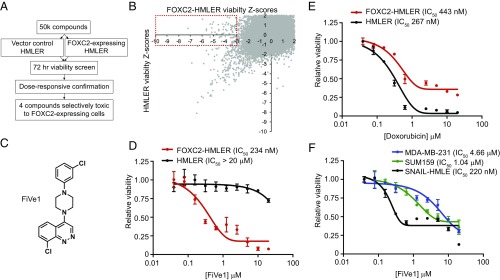
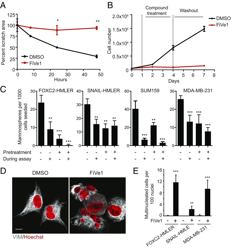
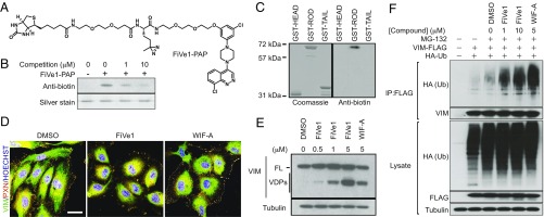
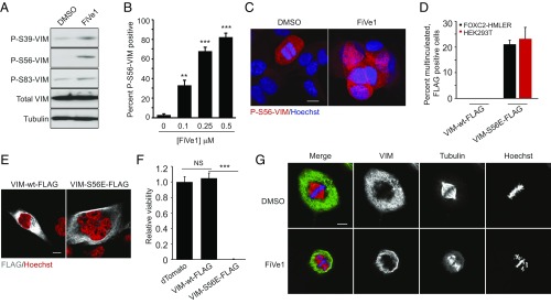
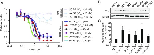
Expression of the transcription factor FOXC2 is induced and necessary for successful epithelial-mesenchymal transition, a developmental program that when activated in cancer endows cells with metastatic potential and the properties of stem cells. As such, identifying agents that inhibit the growth of FOXC2-transformed cells represents an attractive approach to inhibit chemotherapy resistance and metastatic dissemination. From a high throughput synthetic lethal screen, we identified a small molecule, FiVe1, which selectively and irreversibly inhibits the growth of mesenchymally transformed breast cancer cells and soft tissue sarcomas of diverse histological subtypes. FiVe1 targets the intermediate filament and mesenchymal marker vimentin (VIM) in a mode which promotes VIM disorganization and phosphorylation during metaphase, ultimately leading to mitotic catastrophe, multinucleation, and the loss of stemness. These findings illustrate a previously undescribed mechanism for interrupting faithful mitotic progression and may ultimately inform the design of therapies for a broad range of mesenchymal cancers.

Keywords: cancer stem cell; drug discovery; epithelial-to-mesenchymal transition; mitosis; vimentin.


Conflict of interest statement

Conflict of interest statement: M.J.B., S.A.M., P.G.S., and L.L.L. are listed as inventors on a patent application related to the small molecules described in this manuscript.

Figures

Fig. 1.
Fig. 1.
A high throughput screen identifies FiVe1 as a selective inhibitor of FOXC2-expressing breast cancer cells. (A) Schematic representation of the high throughput screening platform used to identify compounds selectively toxic to FOXC2-HMLER cells. (B) Scatterplot of primary screening data displaying plate normalized Z scores of viability for FOXC2-HMLER cells versus control HMLER cells. Contents of red box indicate primary screening hits culled for further analysis. (C) Relative viability measurements of FOXC2 or control HMLER cells exposed to the indicated doses of doxorubicin for 72 h (n = 3, mean and SEM). (D) Structure of FiVe1. (E) Relative viability measurements of FOXC2 and control HMLER cells exposed to the indicated doses of FiVe1 for 72 h (n = 3, mean and SEM). (F) Relative viability measurements of the indicated FOXC2-expressing breast cancer cells after 72-h treatment with FiVe1 (n = 3, mean and SEM).
Fig. 2.
Fig. 2.
FiVe1 irreversibly inhibits stemness-associated properties in mesenchymally transformed breast cancer cells. (A) Quantification of wound closure in an in vitro scratch assay with FOXC2-HMLER cells (n = 3, mean and SD, FiVe1, 1 μM). (B) Growth curves of FOXC2-HMLER cells treated with 1 μM FiVe1 during days 1–4 as noted by dashed vertical lines (n = 3, mean and SD). (C) Quantification of mammospheres from an in vitro mammosphere formation assay with the indicated cell types. Pretreatment indicates a 72-h treatment with 1 μM FiVe1 before plating in mammosphere formation conditions. During assay indicates 1 μM compound treatment during the mammosphere assay (14 d) (n = 3, mean and SD). (D) Representative images of FOXC2-HMLER cells treated for 24 h with DMSO or 500 nM FiVe1 and then stained with DAPI and anti-VIM antibodies. (Scale bar, 10 μm.) (E) Quantification of multinucleation (n > 3 nuclei per cell) in the indicated cell types treated with 500 nM FiVe1 for 24 h (n = 3, mean and SD; *P < 0.05, **P < 0.005, ***P < 0.0005; t test).
Fig. 3.
Fig. 3.
Vimentin is the relevant cellular target of FiVe1. (A) Structure of the photoactivatable affinity probe FiVe1-PAP. (B) Anti-biotin Western blot analysis of recombinant VIM protein exposed to in vitro crosslinking conditions. (FiVe1-PAP, 50 nM; competition, ADP-2341.) (C) Anti-biotin Western blot analysis of the indicated GST-VIM fusion proteins exposed to in vitro crosslinking conditions (FiVe1-PAP, 20 nM). (D) Maximum intensity projections from confocal imaging of vimentin (VIM) and paxillin (PXN) immunostained HUVECs after treatment with 1 μM of the indicated compounds for 1 h. (Scale bar, 20 μm.) (E) Western blot analysis of VIM degradation in FOXC2-HMLER cells after 4 h of treatment with the indicated compounds (FL, full-length VIM; VDPs, VIM degradation products; WIF-A, withaferin A). (F) Western blot analysis of anti-FLAG immunoprecipitated protein content from HEK293T cells transfected with VIM-FLAG and HA-Ub constructs after treatment with the indicated compounds for 4 h.
Fig. 4.
Fig. 4.
Specific engagement of VIM by FiVe1 induces defects in chromosomal alignment during metaphase. (A) Western blotting analysis of phosphorylated VIM protein content from FOXC2-HMLER cells treated for 24 h with FiVe1 (500 nM). Quantification (B) and representative images (C) of immunofluorescent staining for P-S56-VIM from FOXC2-HMLER cells treated with the indicated doses of FiVe1 (n = 3, mean and SD). (D) Quantification of multinucleated, FLAG positive FOXC2-HMLER or HEK293T cells 72 h after transfection with the indicated VIM expression constructs (n = 3, mean and SD). (E) Representative images of FOXC2-HMLER cell immunostained for FLAG protein content 72 h after transfection with the indicated VIM expression constructs. (Scale bar, 10 μm.) (F) Relative viability measurements of FOXC2-HMLER cells 7 d after transduction with lentiviruses encoding the indicated overexpressed transgene products (n = 8, mean and SD). (G) Representative confocal images of thymidine-synced FOXC2-HMLER cells at metaphase immunostained for VIM and β-tubulin (TUB) (FiVe1, 250 nM). (Scale bar, 10 μm.) (**P < 0.005, ***P < 0.0005; NS, not significant; t test).
Fig. 5.
Fig. 5.
FiVe1 inhibits the growth of soft tissue sarcoma (STS) cell lines. (A) Relative proliferation measurements of the indicated VIM negative and STS cell lines treated with a concentration response of FiVe1 (n = 3, mean and SEM). (B) Western blotting analysis for VIM protein levels across the STS panel (FOXC2 = FOXC2-HMLER). (C) Multinucleation analysis of the indicated STS lines treated for 24 h with 1 μM FiVe1 (n = 3, mean and SD; *P < 0.05, t test).

References

    1. Mani SA, et al. The epithelial-mesenchymal transition generates cells with properties of stem cells. Cell. 2008;133:704–715. - PMC - PubMed
    1. May CD, et al. Epithelial-mesenchymal transition and cancer stem cells: A dangerously dynamic duo in breast cancer progression. Breast Cancer Res. 2011;13:202. - PMC - PubMed
    1. Mani SA, et al. Mesenchyme Forkhead 1 (FOXC2) plays a key role in metastasis and is associated with aggressive basal-like breast cancers. Proc Natl Acad Sci USA. 2007;104:10069–10074. - PMC - PubMed
    1. Hollier BG, et al. FOXC2 expression links epithelial-mesenchymal transition and stem cell properties in breast cancer. Cancer Res. 2013;73:1981–1992. - PMC - PubMed
    1. Taube JH, et al. Core epithelial-to-mesenchymal transition interactome gene-expression signature is associated with claudin-low and metaplastic breast cancer subtypes. Proc Natl Acad Sci USA. 2010;107:15449–15454. - PMC - PubMed

Publication types

MeSH terms