

Precancer in ulcerative colitis: the role of the field effect and its clinical implications
- PMID: 29087436
- PMCID: PMC6248676
- DOI: 10.1093/carcin/bgx117
Precancer in ulcerative colitis: the role of the field effect and its clinical implications
Abstract
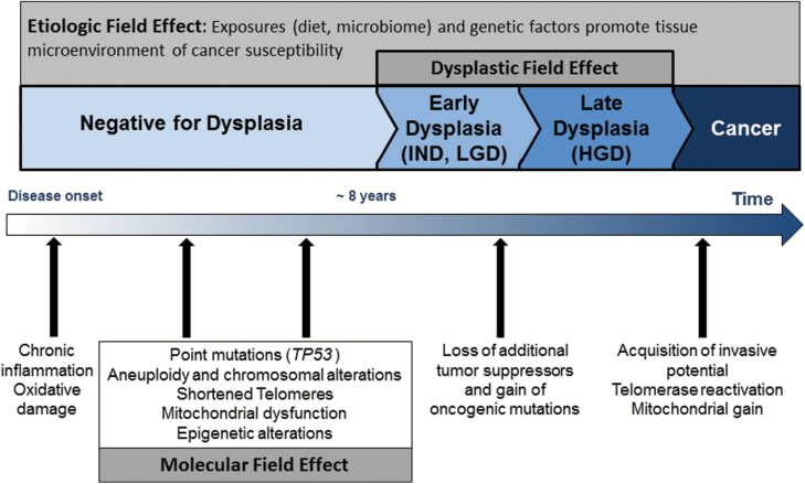
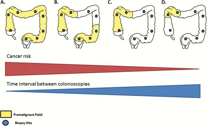
Cumulative evidence indicates that a significant proportion of cancer evolution may occur before the development of histological abnormalities. While recent improvements in DNA sequencing technology have begun to reveal the presence of these early preneoplastic clones, the concept of 'premalignant field' was already introduced by Slaughter more than half a century ago. Also referred to as 'field effect', 'field defect' or 'field cancerization', these terms describe the phenomenon by which molecular alterations develop in normal-appearing tissue and expand to form premalignant patches with the potential to progress to dysplasia and cancer. Field effects have been well-characterized in ulcerative colitis, an inflammatory bowel disease that increases the risk of colorectal cancer. The study of the molecular alterations that define these fields is informative of mechanisms of tumor initiation and progression and has provided potential targets for early cancer detection. Herein, we summarize the current knowledge about the molecular alterations that comprise the field effect in ulcerative colitis and the clinical utility of these fields for cancer screening and prevention.
© The Author(s) 2017. Published by Oxford University Press. All rights reserved. For Permissions, please email: journals.permissions@oup.com.
Figures
References
-
- Nowell P.C. (1976)The clonal evolution of tumor cell populations. Science, 194, 23–28. - PubMed
-
- Martincorena I., et al. (2015)Somatic mutation in cancer and normal cells. Science, 349, 1483–1489. - PubMed
-
- Slaughter D.P., et al. (1953)Field cancerization in oral stratified squamous epithelium; clinical implications of multicentric origin. Cancer, 6, 963–968. - PubMed
Publication types
MeSH terms
Grants and funding
LinkOut - more resources
Full Text Sources
Other Literature Sources
Medical
