

Cumulus cells surrounding oocytes with high developmental competence exhibit down-regulation of phosphoinositol 1,3 kinase/protein kinase B (PI3K/AKT) signalling genes involved in proliferation and survival
- PMID: 29087515
- PMCID: PMC5850344
- DOI: 10.1093/humrep/dex320
Cumulus cells surrounding oocytes with high developmental competence exhibit down-regulation of phosphoinositol 1,3 kinase/protein kinase B (PI3K/AKT) signalling genes involved in proliferation and survival
Abstract
Study question: Is the phosphoinositol 1,3-kinase/protein kinase B (PI3K/AKT) pathway expression profile in cumulus cells (CCs) a potential marker of oocyte competence and predictive of pregnancy outcome?
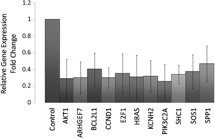
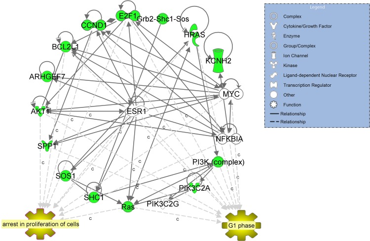
Summary answer: Eleven genes (AKT1, ARHGEF7, BCL2L1, CCND1, E2F1, HRAS, KCNH2, PIK3C2A, SHC1, SOS1 and SPP1) in the PI3K/AKT pathway were significantly down-regulated in CCs from oocytes that went on to produce a pregnancy compared to CCs associated with a negative outcome.
What is known already: The PI3K/AKT pathway plays a pivotal role in the interdependence and continuous feedback between the oocyte and CCs.
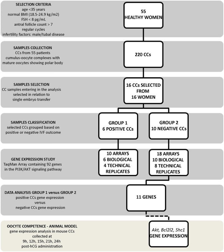
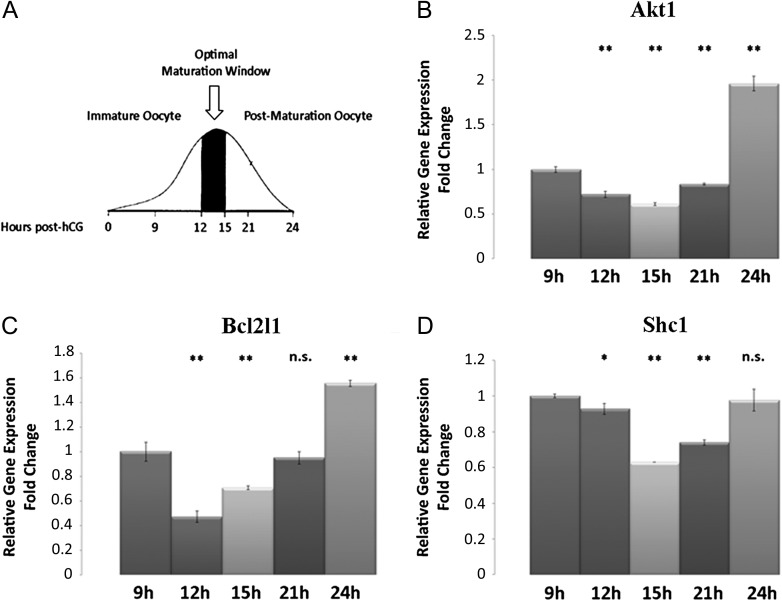
Study design size, duration: The expression analysis of 92 transcripts in the PI3K/AKT pathway in CCs from patients with negative or positive pregnancy outcome, after single embryo transfer, was performed. Mouse CCs target gene expression was conducted to associate the expression profile of PI3K/AKT pathway to oocyte developmental profile.
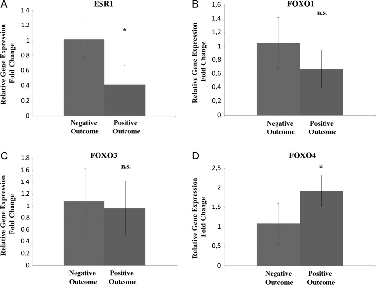
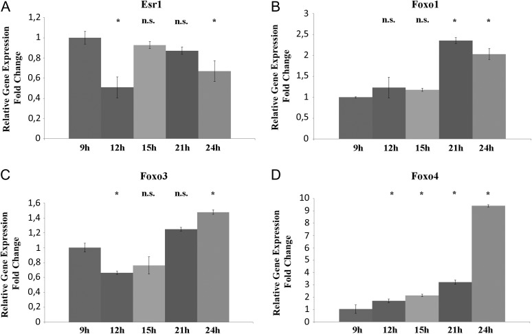
Participants/materials, setting, methods: Fifty-five good prognosis IVF patients who had been referred to IVF or intracytoplasmic sperm injection treatment for male-factor infertility or tubal disease were enroled. CCs from single cumulus-oocyte complexes (COCs) from 16 patients who underwent a single embryo transfer were analyzed. Twenty-five CD-1 mice were used to assess gene expression in CCs associated with oocytes with different competence in relation to hCG priming. A total 220 human COCs were collected. The RNA extracted from CCs of 16 selected patients was used to analyze PI3K/AKT pathway gene expression employing a 96-well custom TaqMan Array. Expression data of CCs associated to positive IVF outcome were compared to data from negative outcome samples. Mice were sacrificed after 9, 12, 15, 21 and 24 h post-hCG administration to obtain CCs from MII oocytes with different developmental competence. Akt1, Bcl2l2 and Shc1 expression were tested in the collected mouse CCs. In addition, the expression of upstream regulator ESR1, the gene encoding for the oestrogen receptor ERβ, and the downstream effectors of the pathway FOXO1, FOXO3 and FOXO4 was evaluated in human and mouse samples.
Main results and the role of chance: Transcripts involved in the PI3K Signaling Pathway were selectively modulated according to the IVF/ICSI outcome of the oocyte. Eleven transcripts in this pathway were significantly down-regulated in all samples of CCs from oocytes with positive when compared those with a negative outcome. These outcomes were confirmed in mouse CCs associated with oocytes at different maturation stages. Expression data revealed that the down-regulation of ESR1 could be related to oocyte competence and is likely to be the driver of expression changes highlighted in the PI3K/AKT pathway.
Limitations reasons for caution: Small sample size and retrospective design.
Wider implications of the findings: The CCs expression profile of PI3K/AKT signaling genes, disclosed a specific CCs gene signature related to oocyte competence. It could be speculated that CCs associated with competent oocytes have completed their role in sustaining oocyte development and are influencing their fate in response to metabolic and hormonal changes by de-activating anti-apoptotic signals.
Study funding/competing interest(s): Supported by Merck Serono an affiliate of Merck KGaA, Darmstadt, Germany (research grant for the laboratory session; Merck KGaA reviewed the manuscript for medical accuracy only before journal submission. The authors are fully responsible for the content of this manuscript, and the views and opinions described in the publication reflect solely those of the authors). The authors declare no conflict of interest.
Keywords: Oocyte competence; PI3K/AKT; cumulus cells; embryo; gene expression; pregnancy outcome.
© The Author 2017. Published by Oxford University Press on behalf of the European Society of Human Reproduction and Embryology.
Figures
References
-
- Afrasiabi E, Hietamäki M, Viitanen T, Sukumaran P, Bergelin N, Törnquist K. Expression and significance of HERG (KCNH2) potassium channels in the regulation of MDA-MB-435S melanoma cell proliferation and migration. Cell Signal 2010;22:57–64. - PubMed
-
- Assou S, Haouzi D, De Vos J, Hamamah S. Human cumulus cells as biomarkers for embryo and pregnancy outcomes. Mol Hum Reprod 2010;16:531–538. - PubMed
MeSH terms
Substances
LinkOut - more resources
Full Text Sources
Other Literature Sources
Research Materials
Miscellaneous
