

Grassystatins D-F, Potent Aspartic Protease Inhibitors from Marine Cyanobacteria as Potential Antimetastatic Agents Targeting Invasive Breast Cancer
- PMID: 29087712
- PMCID: PMC5764543
- DOI: 10.1021/acs.jnatprod.7b00551
Grassystatins D-F, Potent Aspartic Protease Inhibitors from Marine Cyanobacteria as Potential Antimetastatic Agents Targeting Invasive Breast Cancer
Abstract
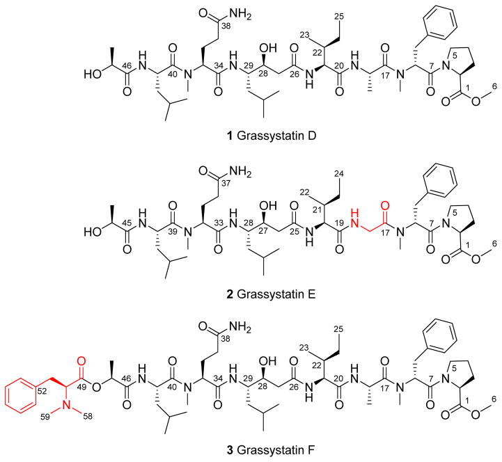
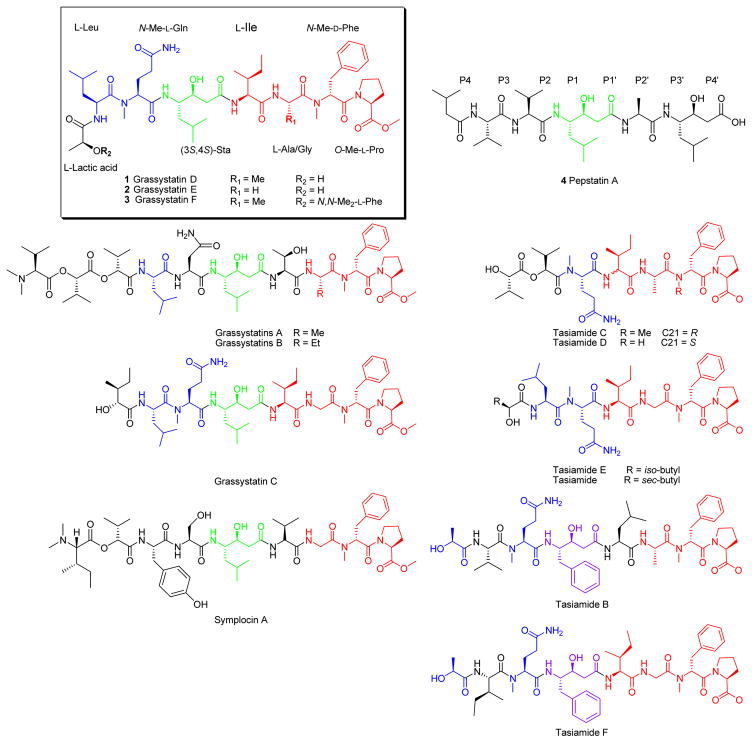
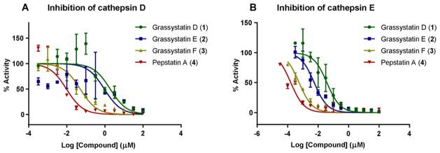
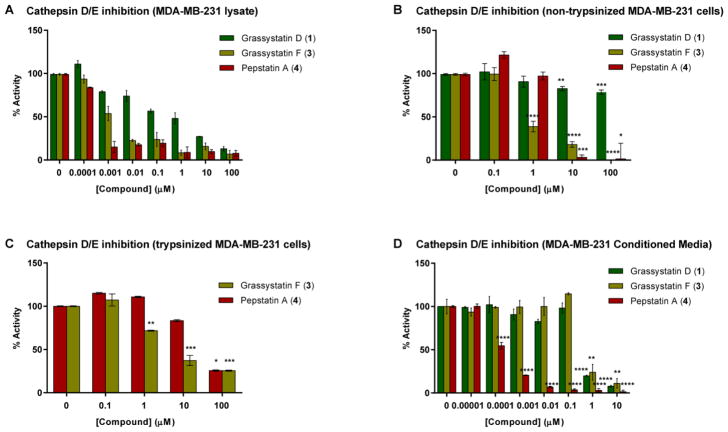
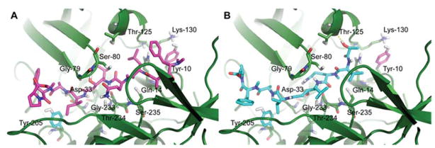
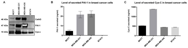
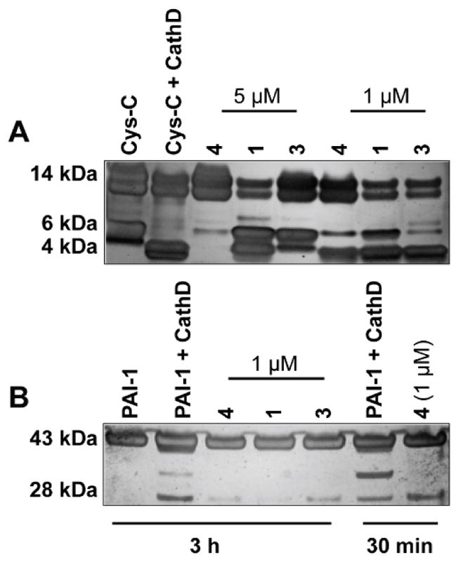
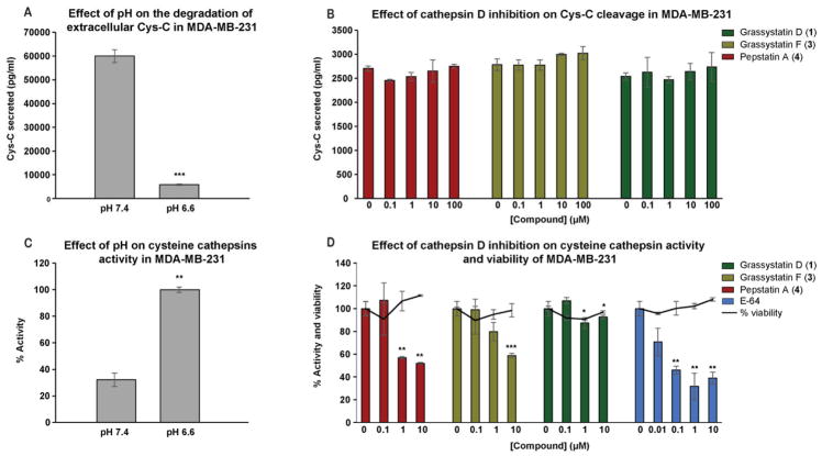
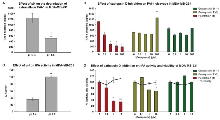
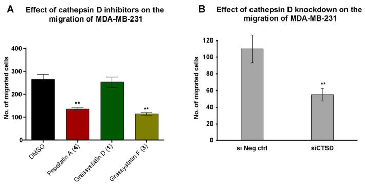
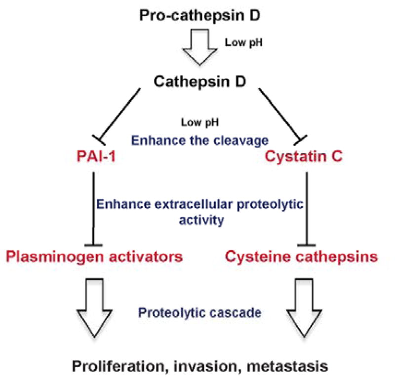
Three new modified peptides named grassystatins D-F (1-3) were discovered from a marine cyanobacterium from Guam. Their structures were elucidated using NMR spectroscopy and mass spectrometry. The hallmark structural feature in the peptides is a statine unit, which contributes to their aspartic protease inhibitory activity preferentially targeting cathepsins D and E. Grassystatin F (3) was the most potent analogue, with IC50 values of 50 and 0.5 nM against cathepsins D and E, respectively. The acidic tumor microenvironment is known to increase the activation of some of the lysosomal proteases associated with tumor metastasis such as cathepsins. Because cathepsin D is a biomarker in aggressive forms of breast cancer and linked to poor prognosis, the effects of cathepsin D inhibition by 1 and 3 on the downstream cellular substrates cystatin C and PAI-1 were investigated. Furthermore, the functional relevance of targeting cathepsin D substrates was evaluated by examining the effect of 1 and 3 on the migration of MDA-MD-231 cells. Grassystatin F (3) inhibited the cleavage of cystatin C and PAI-1, the activities of their downstream targets cysteine cathepsins and tPA, and the migration of the highly aggressive triple negative breast cancer cells, phenocopying the effect of siRNA-mediated knockdown of cathepsin D.
Figures
Similar articles
-
Tasiamide F, a potent inhibitor of cathepsins D and E from a marine cyanobacterium.Bioorg Med Chem. 2016 Aug 1;24(15):3276-82. doi: 10.1016/j.bmc.2016.04.062. Epub 2016 Apr 30. Bioorg Med Chem. 2016. PMID: 27211244 Free PMC article.
-
Grassystatins A-C from marine cyanobacteria, potent cathepsin E inhibitors that reduce antigen presentation.J Med Chem. 2009 Sep 24;52(18):5732-47. doi: 10.1021/jm9009394. J Med Chem. 2009. PMID: 19715320 Free PMC article.
-
Grassystatin-derived peptides selectively inhibit cathepsin E and have low affinity to cathepsin D.Biochem Biophys Res Commun. 2020 Jun 18;527(1):238-241. doi: 10.1016/j.bbrc.2020.04.070. Epub 2020 May 1. Biochem Biophys Res Commun. 2020. PMID: 32446374
-
Proteases as prognostic markers in cancer.Clin Cancer Res. 1996 Apr;2(4):613-8. Clin Cancer Res. 1996. PMID: 9816210 Review.
-
Structures and functions of lysosomal thiol proteinases and their endogenous inhibitor.Curr Top Cell Regul. 1983;22:71-101. doi: 10.1016/b978-0-12-152822-5.50007-5. Curr Top Cell Regul. 1983. PMID: 6347528 Review. No abstract available.
Cited by
-
Chemical diversity of cyanobacterial natural products.Nat Prod Rep. 2025 Jan 22;42(1):6-49. doi: 10.1039/d4np00040d. Nat Prod Rep. 2025. PMID: 39540765 Review.
-
Marine Cyanobacteria: A Source of Lead Compounds and their Clinically-Relevant Molecular Targets.Molecules. 2020 May 8;25(9):2197. doi: 10.3390/molecules25092197. Molecules. 2020. PMID: 32397127 Free PMC article. Review.
-
Total Syntheses of Cathepsin D Inhibitory Izenamides A, B, and C and Structural Confirmation of Izenamide B.Molecules. 2019 Sep 20;24(19):3424. doi: 10.3390/molecules24193424. Molecules. 2019. PMID: 31547147 Free PMC article.
-
Microcystin-LR-Induced Interaction between M2 Tumor-Associated Macrophage and Colorectal Cancer Cell Promotes Colorectal Cancer Cell Migration through Regulating the Expression of TGF-β1 and CST3.Int J Mol Sci. 2023 Jun 23;24(13):10527. doi: 10.3390/ijms241310527. Int J Mol Sci. 2023. PMID: 37445705 Free PMC article.
-
Natural Products from Cyanobacteria: Focus on Beneficial Activities.Mar Drugs. 2019 May 30;17(6):320. doi: 10.3390/md17060320. Mar Drugs. 2019. PMID: 31151260 Free PMC article. Review.
References
-
- Turk B. Nat Rev Drug Discov. 2006;5:785–799. - PubMed
Publication types
MeSH terms
Substances
Grants and funding
LinkOut - more resources
Full Text Sources
Other Literature Sources
Molecular Biology Databases
Miscellaneous
