. 2017 Nov 21;114(47):12501-12506.
doi: 10.1073/pnas.1706855114. Epub 2017 Oct 31.

Efficient gene knockin in axolotl and its use to test the role of satellite cells in limb regeneration

Affiliations

Efficient gene knockin in axolotl and its use to test the role of satellite cells in limb regeneration

Ji-Feng Fei et al. Proc Natl Acad Sci U S A. .

Abstract

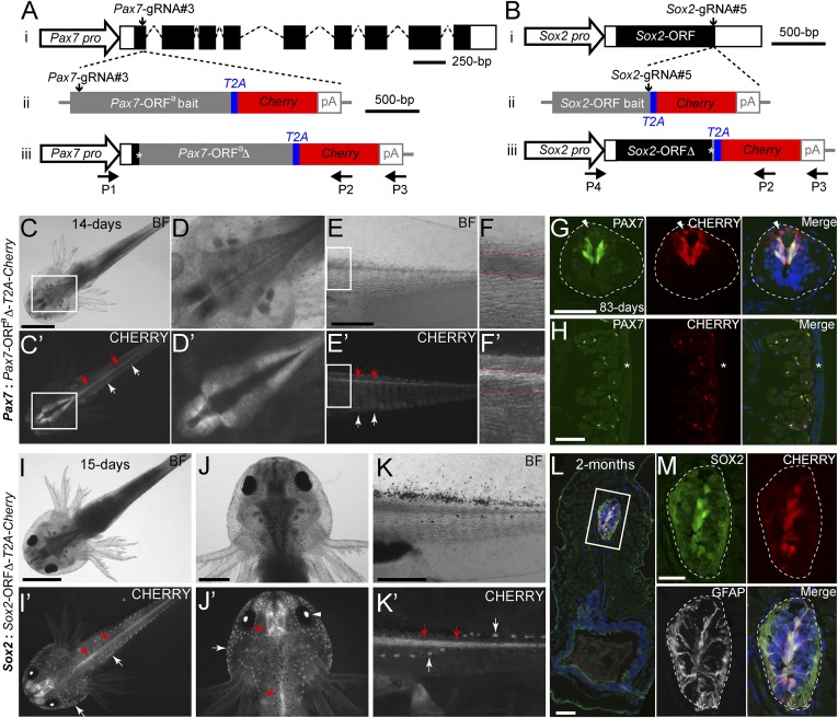
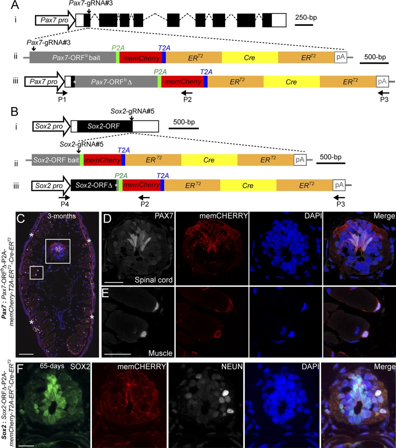
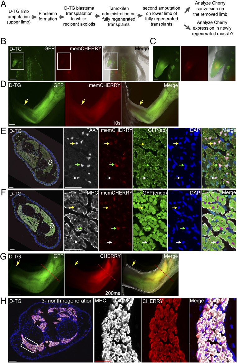
Salamanders exhibit extensive regenerative capacities and serve as a unique model in regeneration research. However, due to the lack of targeted gene knockin approaches, it has been difficult to label and manipulate some of the cell populations that are crucial for understanding the mechanisms underlying regeneration. Here we have established highly efficient gene knockin approaches in the axolotl (Ambystoma mexicanum) based on the CRISPR/Cas9 technology. Using a homology-independent method, we successfully inserted both the Cherry reporter gene and a larger membrane-tagged Cherry-ERT2-Cre-ERT2 (∼5-kb) cassette into axolotl Sox2 and Pax7 genomic loci. Depending on the size of the DNA fragments for integration, 5-15% of the F0 transgenic axolotl are positive for the transgene. Using these techniques, we have labeled and traced the PAX7-positive satellite cells as a major source contributing to myogenesis during axolotl limb regeneration. Our work brings a key genetic tool to molecular and cellular studies of axolotl regeneration.

Keywords: CRISPR/Cas9; knockin; neural stem cells; regeneration; satellite cells.


Conflict of interest statement

The authors declare no conflict of interest.

Figures

Fig. 1.
Fig. 1.
Knockin of a Cherry reporter gene into two axolotl genomic loci through CRISPR/Cas9- mediated homologous-independent integration. (A and B) The knockin strategies for the generation of the Pax7: Pax7-ORFa∆-T2A-Cherry (A) and Sox2: Sox2-ORF∆-T2A-Cherry (B) axolotl reporter lines. (i) The wild-type axolotl Pax7 (A, Dataset S1) and Sox2 (B) gene structures. Solid rectangles represent coding sequences of the exons; empty rectangles, untranslated regions; dashed lines: introns. The gRNA targeting sites are indicated by vertical arrows. (ii) The targeting constructs, pGEMT-Pax7-ORFa-T2A-Cherry-PA (A) and pGEMT-Sox2-ORF-T2A-Cherry-PA (B), contain the entire axolotl Pax7 (A; Pax7-ORFa bait) or Sox2 (B; Sox2-ORF bait) ORF of the cDNA (with the start codon, but without the stop codon) as the bait sequence, followed by T2A, the Cherry coding sequence, and the polyadenylation signal (pA). Vertical arrows indicate the gRNA targeting sites. (iii) The Pax7 (A) and Sox2 (B) alleles after knockin of the Cherry reporter gene. Asterisks indicate the junctions after the integration of the targeting constructs. The newly formed mosaic Pax7 (A) and Sox2 (B) coding sequences are designated Pax7-ORFaΔ and Sox2-ORFΔ, respectively. The horizontal arrows indicate the binding positions of the genotyping primers (P1–P4). (CF′) Bright-field (BF; Upper) and CHERRY fluorescence (Lower) images of 14-d-old Pax7: Pax7-ORFaΔ-T2A-Cherry knockin F0 axolotls. The dorsal (CD′) and lateral (EF′) view images highlight the CHERRY expression in the brain (CD′), spinal cord (red arrows), and trunk muscle (white arrows) compartments. The areas depicted by rectangles in C and C′ and E and E′ are shown at higher magnification in D and D′ and F and F′, respectively. The red dashed lines (F and F′) define the spinal cord region showing that the CHERRY expression is restricted in the dorsal domain of the spinal cord. (Scale bars: 2 mm in C, 1 mm in E.) (G and H) Immunofluorescence for PAX7 (green), CHERRY fluorescence (red), and DAPI (blue) on 10-μm tail cross-cryosections of 83-d-old Pax7: Pax7-ORFaΔ-T2A-Cherry knockin F0 axolotls shows that CHERRY expression is restricted to the PAX7-expressing domain in dorsal spinal cord (G, dashed line circles) and satellite cells (H). The arrowhead indicates inherited CHERRY in a PAX7-negative newborn neuron. The asterisk indicates tail skin. (Scale bars: 100 μm in G and H). (IK′) Bright-field (BF, Upper) and CHERRY fluorescence (Lower) images of 15-d-old Sox2: Sox2-ORFΔ-T2A-Cherry knockin F0 axolotls. The dorsal view (IJ′) and lateral view (K and K′) images highlight the CHERRY expression in the central nervous system (red arrows), the lens (arrowhead), and the lateral line neuromasts (white arrows). (Scale bars: 2 mm in I, 1 mm in J and K.) (L and M) Immunofluorescence for SOX2 (green), GFAP (white), and CHERRY fluorescence (red) combined with DAPI (blue) on 10-μm cross-cryosections of 2-mo-old Sox2: Sox2-ORFΔ-T2A-Cherry knockin F0 axolotls shows that CHERRY expression is restricted to SOX2 positive cells in the spinal cord (dashed circles) (M). GFAP is shown to highlight the morphology of the luminal SOX2-positive radial glial cells expressing GFAP. The rectangle depicted in L is shown as separated or merged images at higher magnification in M. (Scale bars: 100 μm in L, 50 μm in M.)
Fig. 2.
Fig. 2.
Knockin of a large gene cassette into two axolotl genomic loci through a CRISPR/Cas9- mediated homologous-independent approach. (A and B) The knockin strategies for the generation of the Pax7: Pax7-ORFb-P2A-memCherry-T2A-ERT2-Cre-ERT2 (A) and Sox2: Sox2-ORF∆-P2A-memCherry-T2A-ERT2-Cre-ERT2 (B) axolotl transgenic lines. (i) The wild-type axolotl Pax7 (A; Dataset S1) and Sox2 (B) gene structures. Solid rectangles represent coding sequences of the exons; empty rectangles, untranslated regions; dashed lines, introns. Vertical arrows indicate the gRNA targeting sites. (ii) The targeting constructs pGEMT-Pax7-ORFb-P2A-memCherry-T2A- ERT2-Cre-ERT2-PA (A) and pGEMT-Sox2-ORF-P2A-memCherry-T2A-ERT2-Cre-ERT2 -PA (B) contain the entire axolotl Pax7 (A; Pax7-ORFb bait) and Sox2 (B; Sox2-ORF bait) ORF of the cDNA as the bait sequences, followed by the P2A, memCherry (a GAP43 membrane-localization sequence-tagged Cherry gene; Dataset S2), T2A, the ERT2-Cre-ERT2 coding sequences, and the polyadenylation signal (pA). Vertical arrows indicate the gRNA targeting sites. (iii) The Pax7 (A) and Sox2 (B) alleles after successful knockin of the P2A-memCherry-T2A-ERT2-Cre-ERT2 cassettes. Asterisks indicate the junctions after the integration of the targeting constructs. The newly formed mosaic Pax7 (A) and Sox2 (B) coding sequences were designated Pax7-ORFbΔ and Sox2-ORFΔ, respectively. The horizontal arrows indicate the binding positions of the genotyping primers (P1–P4). (CE) Immunofluorescence for PAX7 (white) and memCHERRY (red) combined with DAPI (blue) on 10-μm tail cross-cryosections of 3-mo-old Pax7: Pax7-ORFbΔ-P2A-memCherry-T2A-ERT2-Cre-ERT2 knockin F0 axolotls shows a membrane-juxtaposed CHERRY signal surrounding PAX7-positive radial glial cells in the spinal cord (C and D) and satellite cells (C and E). The rectangles depicted in C are shown as separated or merged images at higher magnification in D and E. Asterisks in C indicate nonspecific CHERRY staining in the dermis, which is also present in the control (SI Appendix, Fig. S6D). (Scale bars: 200 μm in C, 50 μm in D and E.) (F) Immunofluorescence for SOX2 (green), CHERRY (red, memCHERRY), NEUN (white, to label neurons), and DAPI (blue) on 10-μm cross-cryosections of 65-d-old Sox2: Sox2-ORFΔ-P2A-memCherry-T2A-ERT2-Cre-ERT2 knockin F0 axolotls shows a membrane-juxtaposed CHERRY signal surrounding SOX2-positive radial glial cells in the spinal cord. (Scale bar: 50 μm.)
Fig. 3.
Fig. 3.
Lineage tracing of satellite cells during axolotl limb regeneration. (A) Scheme of the satellite cell lineage tracing experiment. (BD) Live images of double-transgenic limbs after blastema transplantation and before tamoxifen treatment (n = 6). The GFP, CHERRY fluorescence, and merged (with bright-field) images of the transplanted limb blastema from the double-transgenic (D-TG) (SI Appendix, Fig. S7) donor to a white recipient at 2 wk (B and C) and 3 mo (D) post-transplantation. Note that the membrane-CHERRY signal is very dim in satellite cells in live imaging. Rectangular regions in B are shown at higher magnification in C. (Scale bars: 1 mm in B and D, 500 μm in C.) (E and F) Cross-section of limbs at 2 wk after tamoxifen treatment. Immunofluorescence for PAX7 (white, E) or MHC (white, F), CHERRY (red, memCHERRY), GFP [green, from antibody (ab) staining, in E] or endogenous (endo) GFP fluorescence (green, in F) combined with DAPI (blue) on adjacent 10-μm limb cross-cryosections (showing limb muscle) (n = 3). Green and yellow arrowheads indicate strong and dim cytoplasmic CHERRY-expressing (converted) cells, respectively. White arrowhead marks unconverted, memCHERRY-expressing cells. Muscle fibers (MHC-labeled) in the converted, mature limb remain CHERRY-negative. In E and F, the areas depicted by the rectangle in the leftmost panel are shown at higher magnification at the three right panels (Scale bars: 200 μm in left and 50 μm in right.) (G) Live images of tamoxifen-converted limbs after completion of regeneration: GFP, CHERRY fluorescence, and merged (with bright-field) images (n = 3). Arrows indicate the first amputation plane from the transplantation, and the red dashed line shows the plane of amputation after tamoxifen conversion. Exposure time for CHERRY, 200 ms. Note that Cherry driven by the CAGGS promoter is more strongly expressed in muscle tissues compared with satellite cells (10 s in Fig. 3D vs. 200 ms in Fig. 3G). Note that the GFP signal observed in the digit muscles represents GFP expression from incomplete conversion of the tandem array of LoxP reporter cassettes found in this reporter strain (SI Appendix, Fig. S10). (Scale bar: 1 mm.) (H) CHERRY-expressing satellite cells contribute to muscle regeneration. Cross-section of the limb that regenerated after induction of CHERRY expression from the LoxP reporter in PAX7-positive cells. Immunofluorescence for MHC (white) and CHERRY (red) combined with DAPI (blue) of a 3-mo regenerated limb showing robust expression of cytoplasmic CHERRY in muscle cells (n = 3). The area depicted by the rectangle in the leftmost panel is shown at higher magnification in the three right panels. (Scale bars: 200 μm in left and 50 μm in right.)

References

    1. Kragl M, et al. Cells keep a memory of their tissue origin during axolotl limb regeneration. Nature. 2009;460:60–65. - PubMed
    1. Sandoval-Guzmán T, et al. Fundamental differences in dedifferentiation and stem cell recruitment during skeletal muscle regeneration in two salamander species. Cell Stem Cell. 2014;14:174–187. - PubMed
    1. Khattak S, et al. Germline transgenic methods for tracking cells and testing gene function during regeneration in the axolotl. Stem Cell Rep. 2013;1:90–103. - PMC - PubMed
    1. Nakade S, et al. Microhomology-mediated end-joining-dependent integration of donor DNA in cells and animals using TALENs and CRISPR/Cas9. Nat Commun. 2014;5:5560. - PMC - PubMed
    1. Shi Z, et al. Heritable CRISPR/Cas9-mediated targeted integration in Xenopus tropicalis. FASEB J. 2015;29:4914–4923. - PubMed

Publication types

MeSH terms

Associated data