

Assessing for Cardiotoxicity from Metal-on-Metal Hip Implants with Advanced Multimodality Imaging Techniques
- PMID: 29088037
- PMCID: PMC6948834
- DOI: 10.2106/JBJS.16.00743
Assessing for Cardiotoxicity from Metal-on-Metal Hip Implants with Advanced Multimodality Imaging Techniques
Abstract
Background: High failure rates of metal-on-metal (MoM) hip implants prompted regulatory authorities to issue worldwide safety alerts. Circulating cobalt from these implants causes rare but fatal autopsy-diagnosed cardiotoxicity. There is concern that milder cardiotoxicity may be common and underrecognized. Although blood metal ion levels are easily measured and can be used to track local toxicity, there are no noninvasive tests for organ deposition. We sought to detect correlation between blood metal ions and a comprehensive panel of established markers of early cardiotoxicity.
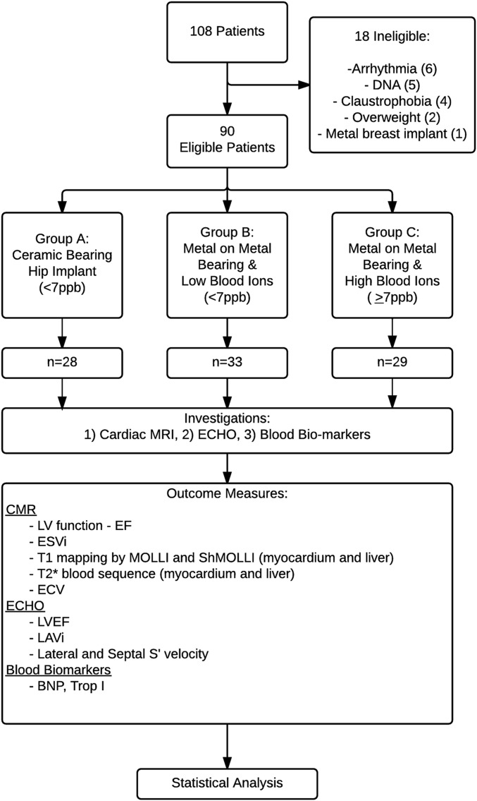
Methods: Ninety patients were recruited into this prospective single-center blinded study. Patients were divided into 3 age and sex-matched groups according to implant type and whole-blood metal ion levels. Group-A patients had a ceramic-on-ceramic [CoC] bearing; Group B, an MoM bearing and low blood metal ion levels; and Group C, an MoM bearing and high blood metal-ion levels. All patients underwent detailed cardiovascular phenotyping using cardiac magnetic resonance imaging (CMR) with T2*, T1, and extracellular volume mapping; echocardiography; and cardiac blood biomarker sampling. T2* is a novel CMR biomarker of tissue metal loading.
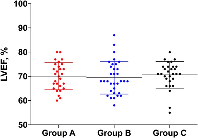
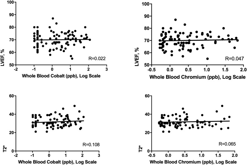
Results: Blood cobalt levels differed significantly among groups A, B, and C (mean and standard deviation [SD], 0.17 ± 0.08, 2.47 ± 1.81, and 30.0 ± 29.1 ppb, respectively) and between group A and groups B and C combined. No significant between-group differences were found in the left atrial or ventricle size, ejection fraction (on CMR or echocardiography), T1 or T2* values, extracellular volume, B-type natriuretic peptide level, or troponin level, and all values were within normal ranges. There was no relationship between cobalt levels and ejection fraction (R = 0.022, 95% confidence interval [CI] = -0.185 to 0.229) or T2* values (R = 0.108, 95% CI = -0.105 to 0.312).
Conclusions: Using the best available technologies, we did not find that high (but not extreme) blood cobalt and chromium levels had any significant cardiotoxic effect on patients with an MoM hip implant. There were negligible-to-weak correlations between elevated blood metal ion levels and ejection fraction even at the extremes of the 95% CI, which excludes any clinically important association.
Level of evidence: Therapeutic Level II. See Instructions for Authors for a complete description of levels of evidence.
Figures
References
-
- Bozic KJ, Kurtz S, Lau E, Ong K, Chiu V, Vail TP, Rubash HE, Berry DJ. The epidemiology of bearing surface usage in total hip arthroplasty in the United States. J Bone Joint Surg Am. 2009. July;91(7):1614-20. - PubMed
-
- Hart AJ, Sabah S, Henckel J, Lewis A, Cobb J, Sampson B, Mitchell A, Skinner JA. The painful metal-on-metal hip resurfacing. J Bone Joint Surg Br. 2009. June;91(6):738-44. - PubMed
-
- Pandit H, Glyn-Jones S, McLardy-Smith P, Gundle R, Whitwell D, Gibbons CL, Ostlere S, Athanasou N, Gill HS, Murray DW. Pseudotumours associated with metal-on-metal hip resurfacings. J Bone Joint Surg Br. 2008. July;90(7):847-51. - PubMed
-
- Schaffer AW, Pilger A, Engelhardt C, Zweymueller K, Ruediger HW. Increased blood cobalt and chromium after total hip replacement. J Toxicol Clin Toxicol. 1999;37(7):839-44. - PubMed
-
- Case CP, Langkamer VG, James C, Palmer MR, Kemp AJ, Heap PF, Solomon L. Widespread dissemination of metal debris from implants. J Bone Joint Surg Br. 1994. September;76(5):701-12. - PubMed
MeSH terms
Substances
LinkOut - more resources
Full Text Sources
Other Literature Sources
Medical
Research Materials
