

Melanosis coli: Harmless pigmentation? A case-control retrospective study of 657 cases
- PMID: 29088250
- PMCID: PMC5663380
- DOI: 10.1371/journal.pone.0186668
Melanosis coli: Harmless pigmentation? A case-control retrospective study of 657 cases
Abstract
Backgrounds and aims: The association of melanosis coli with the development of colorectal polyps remains uncertain.
Methods: From a total of 18263 patients who had received colonoscopy in our hospital, 219 with melanosis coli cases and 438 controls matched by age and sex (at 1:2 ratio) were included in this study. The association of incidence, number, location, and pathology of colorectal neoplasm with grades and distribution of melanosis coli were analyzed.
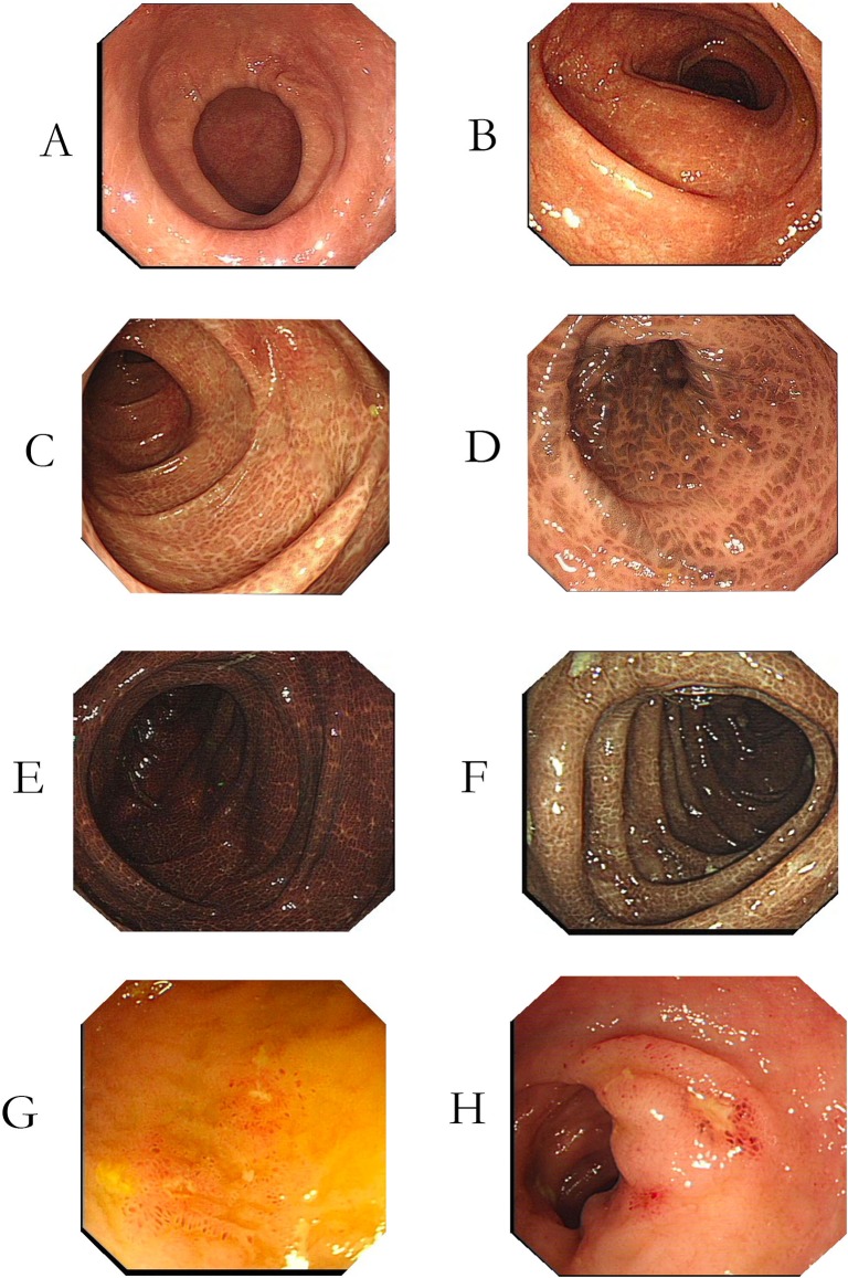
Results: Melanosis coli was associated with significantly more colorectal polyps than control, a higher incidence of numerous colorectal polyps (number ≥ 20) (7.3% vs 0.5%; p < 0.001), and higher number of small colorectal polyps (diameter ≤ 5 mm; p < 0.01). Patients with melanosis coli had higher incidences of low-grade adenomas (31.1% vs 23.3%, p < 0.05) and non-adenoma polyps (20.1% vs 12.8%, p < 0.05) than the controls. On multivariate analysis, melanosis coli was independently associated with increased detecting rates of low grade adenoma (OR = 1.54; 95%: 1.06-2.23; p < .05), non-adenoma polyp (OR = 1.72; 95%: 1.11-2.70; p < .05) and numerous polyps (OR = 16.2, 95%: 3.66-71.6; p < .05). There was no significant difference in the incidence of high-grade adenomas or adenocarcinomas in the two population groups, but the numbers of these lesions were insufficient to permit firm conclusions. No significant differences in incidence, number, and pathology of colorectal polyps between individuals with melanosis coli of three different grades of severity were found. Melanosis located predominantly in the right colon had an interestingly lower incidence of colonic polyps in right colon than did melanosis located predominantly in the left colon or total colon (8.9% vs. 26.3%, 24.0%, p < 0.05). Patients with melanosis coli had significantly more nonspecific distal ileal ulcers than did controls (8.0% vs 0%, p < 0.001).
Conclusion: Melanosis coli is associated with a higher incidence and number of colonic non-adenoma polyps and low-grade adenomas, and higher incidence of distal ileal ulcers. Melanosis coli may not be a harmless pigmentation, but a sign of chronic injury of colonic and intestinal mucosa.
Conflict of interest statement
Figures
References
-
- Steer HW, Colin-Jones DG. Melanosis coli: studies of the toxic effects of irritant purgatives. J Pathol. 1975;115(4):199–205. doi: 10.1002/path.1711150403 - DOI - PubMed
-
- Ghadially FN, Parry EW. An electron-microscope and histochemical study of melanosis coli. J Pathol Bacteriol. 1966;92(2):313–7. doi: 10.1002/path.1700920207 - DOI - PubMed
-
- Cruveilhier J. Anatomie pathologique du corps humain, ou Descriptions, avec figures lithographie\0301es et colorie\0301es, des diverses alte\0301rations morbides dont le corps humain est susceptible: Paris, 1829–1835; 1835.
-
- Virchow R. Die pathologischen Pigmente. Arch. Pathol. Ant, 1847; 1(2): 379–404. https://doi.org/10.1007/BF01975874 - DOI
MeSH terms
LinkOut - more resources
Full Text Sources
Other Literature Sources
Medical
