

Review
doi: 10.1093/brain/awx270.
A little man of some importance
Affiliations
- PMID: 29088352
- PMCID: PMC5841206
- DOI: 10.1093/brain/awx270
Item in Clipboard
Review
A little man of some importance
Brain.
.
Abstract
Eighty years ago, Penfield and Boldrey introduced the homunculus in a paper published in Brain. In a reappraisal of the iconic aide-mémoire, Marco Catani reanalyses the original data, and argues that through its extended network the homunculus holds the key to the precise coding that results in coordinated activation of peripheral muscles.
Figures
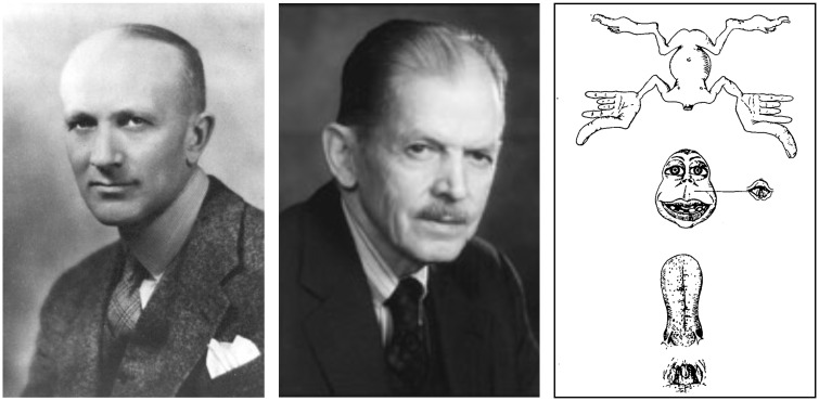
Wilder Penfield (left) and Edwin Boldrey (centre), creators of the homunculus (right).
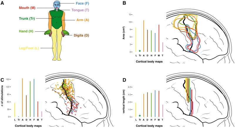
Representation of the motor stimulations for different body parts. (A) Colour-coding of different body parts. (B) Areas of the surface maps enclosing all motor stimulation points for each body part. (C) Count of the number of stimulations and (D) measurements of the vertical length of the surface maps for each body part. Original data are derived from Penfield and Boldrey (1937). The asterisk indicates the impossibility of generating any measurement of the area and length from the few stimulations recorded for the trunk. Similar maps for the somatosensory stimulations are provided in Supplementary Fig. 1.
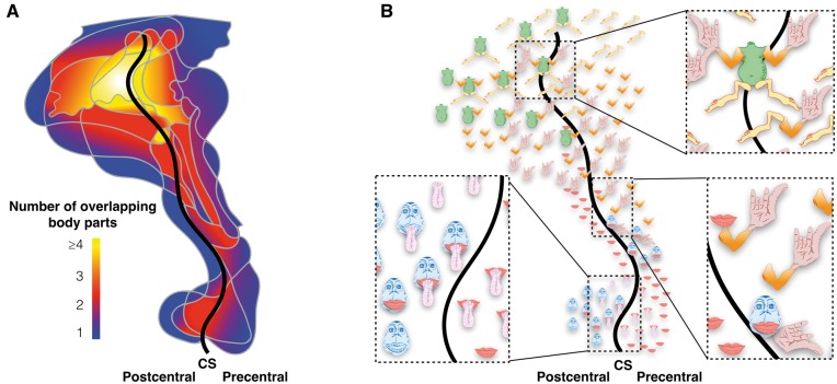
Representation of the overlap between the somatosensory surface maps of different body parts. (A) Heat map of the degree of overlap ranging from blue (one body part) to yellow (four or more body parts). (B) Map indicating the overlapping body parts of the homunculus. CS = central sulcus. Original data are derived from Penfield and Boldrey (1937).
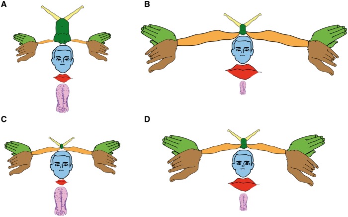
The motor-sensory homunculus redrawn. (A) The proportions of this first homunculus correspond to those of the original reproduced in Fig. 1. All other homunculi in B–D are derived from an average of the motor and somatosensory maps produced in Fig. 2 and Supplementary Fig. 1. (B) Homunculus generated from the surface maps. (C) Homunculus derived from the vertical length measurements. (D) Homunculus derived from the number of stimulation points. All measurements are from Penfield and Boldrey (1937).
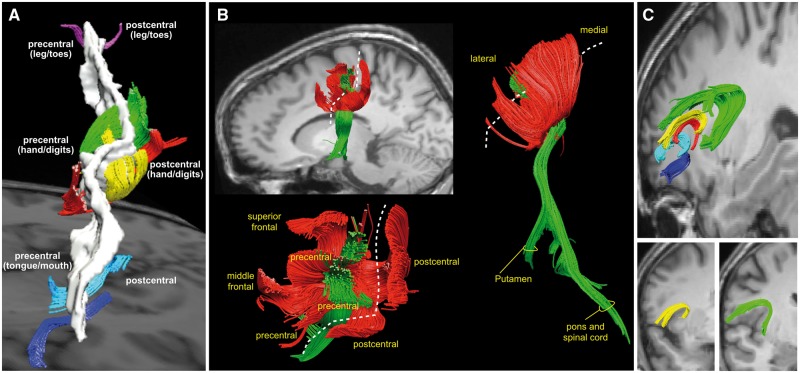
Tractography-based reconstructions of large association and projection tracts of the homuncular cortex. (A) Short association tracts connecting the precentral and postcentral gyri. In the hand knob region, these U-shaped tracts occupy a large volume and show a high degree of complexity (displayed in green, red and yellow colours). In the ventral region of the face and tongue (dark blue and cyan tracts) and dorsal region of the legs and toes (purple tracts) these connections are less prominent. (B) Short association (red) and long projection (green) tracts of the hand knob region from a lateral (upper left), dorsal (lower left) and posterior (right) view. The dashed line indicates the trajectory of the central sulcus. The short association tracts converge to the precentral regions of the hand knob area from the postcentral gyrus and the posterior regions of the superior and middle frontal gyri. The projection tracts are enclosed within the U-shaped tracts and connect the precentral gyrus to the putamen (corticostriatal fibres), the pontine nuclei (corticopontine tracts) and the spinal cord (corticospinal tract). (C) The fronto-insular tracts connect the frontal opercular cortex to the anterior insula. The connections from the precentral and subcentral/postcentral cortex are displayed in yellow and green, respectively. Please note that there is no correspondence between the colours used for these images and the colours in the previous figures. All images modified from Catani et al. (2012).
References
-
- Catani M, Dell'Acqua F, Vergani F, Malik F, Hodge H, Roy P, et al. Short frontal lobe connections of the human brain. Cortex 2012; 48: 273–91. - PubMed
-
- Farrell DF, Burbank N, Lettich E, Ojemann GA. Individual variation in human motor-sensory (rolandic) cortex. J Clin Neurophysiol 2007; 24: 286–93. - PubMed
-
- Foerster O. The motor cortex in man in the light of Hughlings Jackson's doctrines. Brain 1936; 59: 135–59.
Publication types
MeSH terms
Grants and funding
LinkOut - more resources
Full Text Sources
Other Literature Sources
Miscellaneous
