. 2017 Nov 1;140(11):2838-2850.
doi: 10.1093/brain/awx249.

Choline transporter mutations in severe congenital myasthenic syndrome disrupt transporter localization

Affiliations

Choline transporter mutations in severe congenital myasthenic syndrome disrupt transporter localization

Haicui Wang et al. Brain. .

Abstract

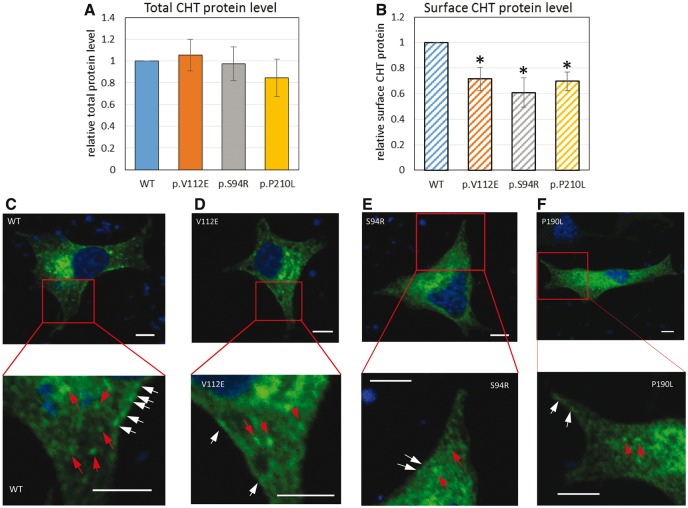
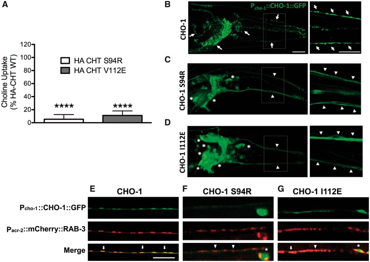
The presynaptic, high-affinity choline transporter is a critical determinant of signalling by the neurotransmitter acetylcholine at both central and peripheral cholinergic synapses, including the neuromuscular junction. Here we describe an autosomal recessive presynaptic congenital myasthenic syndrome presenting with a broad clinical phenotype due to homozygous choline transporter missense mutations. The clinical phenotype ranges from the classical presentation of a congenital myasthenic syndrome in one patient (p.Pro210Leu), to severe neurodevelopmental delay with brain atrophy (p.Ser94Arg) and extend the clinical outcomes to a more severe spectrum with infantile lethality (p.Val112Glu). Cells transfected with mutant transporter construct revealed a virtually complete loss of transport activity that was paralleled by a reduction in transporter cell surface expression. Consistent with these findings, studies to determine the impact of gene mutations on the trafficking of the Caenorhabditis elegans choline transporter orthologue revealed deficits in transporter export to axons and nerve terminals. These findings contrast with our previous findings in autosomal dominant distal hereditary motor neuropathy of a dominant-negative frameshift mutation at the C-terminus of choline transporter that was associated with significantly reduced, but not completely abrogated choline transporter function. Together our findings define divergent neuropathological outcomes arising from different classes of choline transporter mutation with distinct disease processes and modes of inheritance. These findings underscore the essential role played by the choline transporter in sustaining acetylcholine neurotransmission at both central and neuromuscular synapses, with important implications for treatment and drug selection.

Keywords: CHT; CHT trafficking; SLC5A7; choline uptake; congenital myasthenic syndrome.


Figures

Figure 1
Figure 1
Family pedigrees, and mutations detected in SLC5A7. (A–C) Family pedigrees and whole-exome sequencing results. Whole-exome sequencing was done for affected members of each family (Family 1 Patient IV:1, Family 2 Patient V:3 and Family 3 Patient III:5) revealed a single novel deleterious homozygous variant in each subject in the same gene SLC5A7. For Family 3, two siblings (Patients III:3 and III:4) affected with a separate genetic disorder (Fowler syndrome) are marked out in grey. (D) Sequence alignment displays both affected residues (p.Ser94Arg and p.Val112Glu) high evolutionarily sequence conservation while p.Pro210Leu less conserved. (E) CHT topology. The 580 amino acid CHT protein has 13 transmembrane spanning domains and the N-terminus is extracellular while C-terminus is intracellular. The three amino acid substitutions from the missense mutations are indicated in brackets. Two sets of constructs are used in this study, the tags of each set are marked out at the termini.
Figure 2
Figure 2
Recessive CHT missense mutations lead to reduced membrane localization. (A and B) The total and surface N-terminal HA-CHT protein level was assessed by ELISA as described in (Bogatcheva et al., 2007). Protein level is shown in arbitrary units (AU) as a ratio of specific horseradish peroxidase (HRP) activity of mutant to wild-type CHT after anti-HA-HRP antibody binding, (A) total CHT protein level and (B) surface CHT protein level, which is also further normalized to total protein expression. Results are presented as means ± standard error (SE) (n = 3, *P < 0.05, one-way ANOVA). (C–F) CHT cellular localization in PC12 cells transfected with CHTWT-MYC (C), CHTS94R-MYC (D), and CHTV112E-MYC (E), CHTP210L-MYC (F) expression constructs. Transient expressed myc-tagged CHT in transfected PC12 cells was detected via immunostaining using myc antibody (green). Nucleus is stained in DAPI (blue). Scale bar = 5 µm. Bottom panels show magnified view of the red square area of the corresponding top panel. CHTWT-MYC (C) has intracellular vesicular localization (red arrows) and very clear membrane localization (white arrows). CHTV112E-MYC (D) has similar intracellular localization but decreased membrane localization compared to CHTWT-MYC. CHTS94R-MYC (E) and CHTP210L-MYC (F) do not show significant vesicular and membrane localization.
Figure 3
Figure 3
Recessive CHT missense mutation results in altered synaptic localization and have several reduced uptake activity. (A) Choline transport activity is significantly reduced in cells transfected with mutant (p.Val112Glu and p.Ser94Arg) versus wild-type constructs. CHT activity was diminished for both mutants p.Ser94Arg and p.Val112Glu compared to wild-type CHT (HA-CHTS94R: n = 4, *P < 0.0001; HA-CHTV112E: n = 4, *P < 0.0001, one-way ANOVA, Dunnett’s post hoc test). Specific choline uptake was determined by subtracting the uptake obtained in transfected cells with that of mock (vector) transfected cells obtained in assays conducted in parallel. (B) Wild-type Pcho-1::CHO-1::GFP localization and expression in vivo in cholinergic neuron cell bodies and synapses in C. elegans. Right panel: Increased magnification (dotted line box) with arrows indicating normal CHO-1 trafficking as revealed by a punctate labelling patterning in neuronal processes. Scale bar = 20 µm. (C) Expression of CHO-1 Ser94Arg GFP-tagged protein under the expression of the cho-1 native promoter. The mutated protein appears diffuse in neuronal processes and significantly trapped in cell bodies of cholinergic neurons (indicated by asterisks). Right panel: Increased magnification (dotted line box) indicates that mutated CHO-1 does not localize to synaptic regions in neuronal processes. The florescent intensity is ∼3-fold lower than wild-type when injected at similar concentration (adjusted acquisition is shown). (D) Expression of CHO-1 Ile112Glu GFP-tagged protein, under expression of cho-1 native promoter. The mutated protein appears diffuse in neuronal processes and partially trapped in cell bodies of some cholinergic neurons (indicated by asterisks). Right panel: Increased magnification (dotted line box) where mutated CHO-1 does not exhibit GFP puncta in neuronal processes, indicating the absence of proper CHO-1 localization in synaptic vesicles. (E–G) Mutated Pcho-1::CHO-1::GFP localization and expression in vivo in the C. elegans cholinergic motor neurons. (E) Wild-type CHO-1 protein, expressed under the cho-1 native promoter, exhibits normal co-localization with cholinergic neuron synaptic marker, Pacr-2::mCherry::RAB-3 (arrows). Whereas (F) CHO-1 Ser94Arg and (G) CHO-1 Ile112Glu GFP-tagged proteins expressed under the cho-1 native promoter lack the complete synaptic co-localization observed in wild-type protein expression (arrowheads), with CHO-1 Ile112Glu showing occasional correct localization (arrow). CHO-1 Ser94Arg and CHO-1 Ile112Glu mutant proteins appear to be partially trapped in the cell bodies (asterisks).

References

    1. Ahmed MY, Al-Khayat A, Al-Murshedi F, Al-Futaisi A, Chioza BA, Pedro Fernandez-Murray J, et al. A mutation of EPT1 (SELENOI) underlies a new disorder of Kennedy pathway phospholipid biosynthesis. Brain 2017; 140: 547–54. - PMC - PubMed
    1. Apparsundaram S, Ferguson SM, George AL Jr, Blakely RD. Molecular cloning of a human, hemicholinium-3-sensitive choline transporter. Biochem Biophys Res Commun 2000; 276: 862–7. - PubMed
    1. Arredondo J, Lara M, Gospe SM Jr, Mazia CG, Vaccarezza M, Garcia-Erro M, et al. Choline acetyltransferase mutations causing congenital myasthenic syndrome: molecular findings and genotype-phenotype correlations. Hum Mutat 2015; 36: 881–93. - PMC - PubMed
    1. Barwick KE, Wright J, Al-Turki S, McEntagart MM, Nair A, Chioza B, et al. Defective presynaptic choline transport underlies hereditary motor neuropathy. Am J Hum Genet 2012; 91: 1103–7. - PMC - PubMed
    1. Bauché S, O'Regan S, Azuma Y, Laffargue F, McMacken G, Sternberg D, et al. Impaired presynaptic high-affinity choline transporter causes a congenital myasthenic syndrome with episodic apnea. Am J Hum Genet 2016; 99: 753–61. - PMC - PubMed

MeSH terms