. 2017 Nov 9;551(7679):247-250.
doi: 10.1038/nature24297. Epub 2017 Nov 1.

Drug-tolerant persister cancer cells are vulnerable to GPX4 inhibition

Affiliations

Drug-tolerant persister cancer cells are vulnerable to GPX4 inhibition

Matthew J Hangauer et al. Nature. .

Abstract

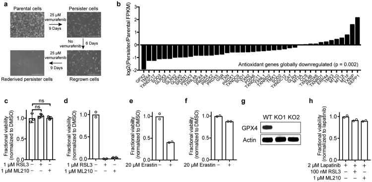
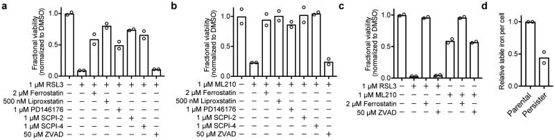
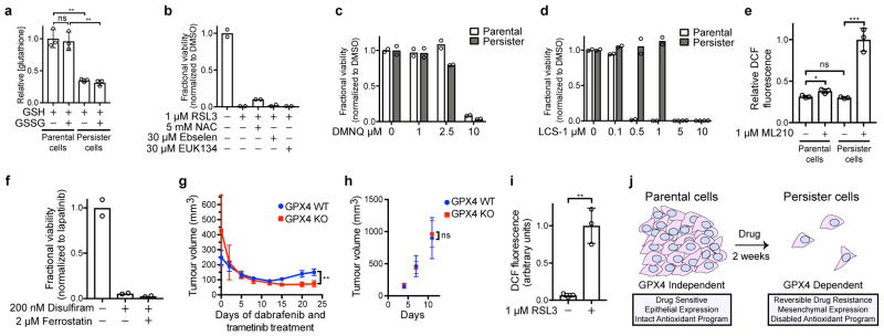
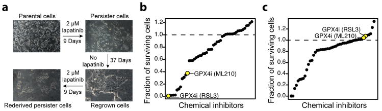
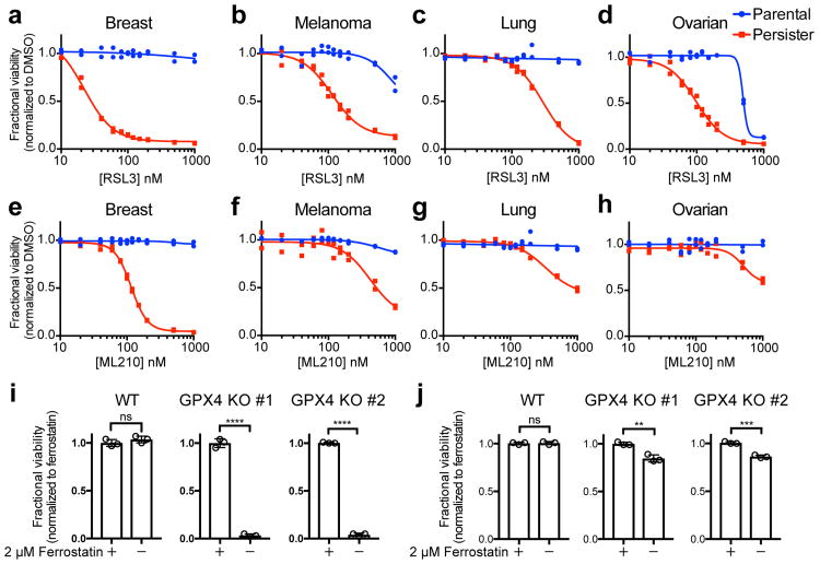
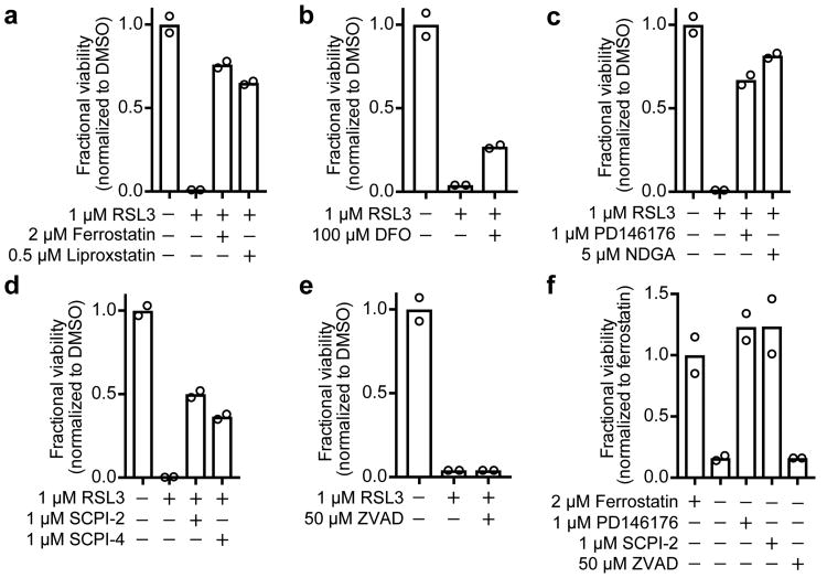
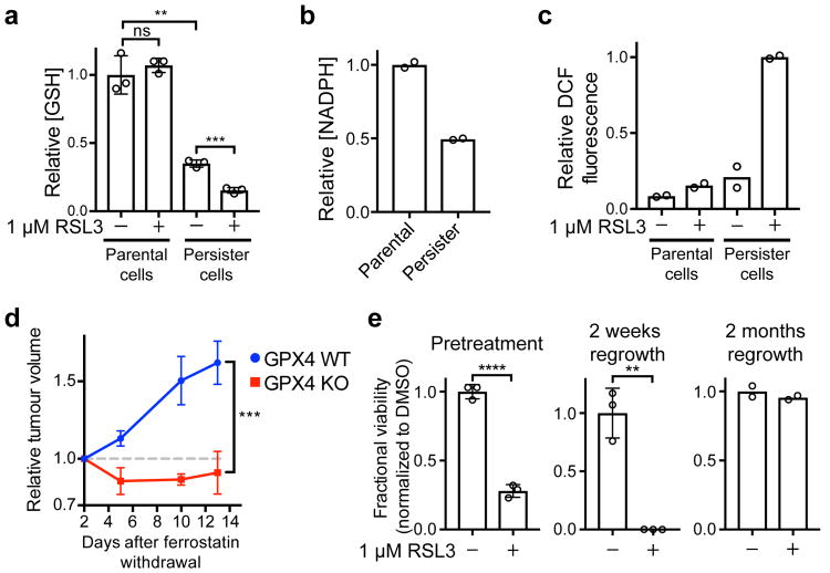
Acquired drug resistance prevents cancer therapies from achieving stable and complete responses. Emerging evidence implicates a key role for non-mutational drug resistance mechanisms underlying the survival of residual cancer 'persister' cells. The persister cell pool constitutes a reservoir from which drug-resistant tumours may emerge. Targeting persister cells therefore presents a therapeutic opportunity to impede tumour relapse. We previously found that cancer cells in a high mesenchymal therapy-resistant cell state are dependent on the lipid hydroperoxidase GPX4 for survival. Here we show that a similar therapy-resistant cell state underlies the behaviour of persister cells derived from a wide range of cancers and drug treatments. Consequently, we demonstrate that persister cells acquire a dependency on GPX4. Loss of GPX4 function results in selective persister cell ferroptotic death in vitro and prevents tumour relapse in mice. These findings suggest that targeting of GPX4 may represent a therapeutic strategy to prevent acquired drug resistance.


Figures

Extended Data Figure 1
Extended Data Figure 1. Additional data demonstrating persister cell ferroptosis sensitivity
a, A375 melanoma persister cells are reversibly drug-resistant. Scale bars indicate 400 μm. b, Global antioxidant gene-expression is downregulated in BT474 persister cells. P value calculated using a two-tailed Wilcoxon signed rank test. c, MCF10A parental cells and d, BT474 persister cells derived from carboplatin and paclitaxel were treated with RSL3 or ML210 for three days. e, BT474 persister cells or f, parental cells were treated with erastin for five days. g, Western blot demonstrating GPX4 knockout in two distinct A375 clones (clone 1, KO1; clone 2, KO2). For gel source data, see Supplementary Figure 1. h, BT474 parental cells co-treated with 2 μM lapatinib and RSL3 or ML210 for three days. Data are plotted as means and error bars represent standard deviation. c, n = 3 and d-h, n = 2 biologically independent samples. c, P value calculated from a two-tailed t test where ns represents P > 0.05. All data are representative of two separate experiments.
Extended Data Figure 2
Extended Data Figure 2. Additional data demonstrating that GPX4 inhibition causes ferroptosis in persister cells
A375 persister cells were treated with a, RSL3 or b, ML210 and ferroptosis rescue compounds for three days. c, PC9 persister cells were treated with RSL3 or ML210 and ferroptosis rescue compounds for three days. d, Relative concentration of total labile iron in BT474 parental and persister cells. Data are plotted as means and error bars represent standard deviation. a-d, n = 2 biologically independent samples. From two-tailed t tests, *, P < 0.05; **, P < 0.01; ***, P < 0.001; ****, P < 0.0001; ns, P > 0.05. All data are representative of two separate experiments.
Extended Data Figure 3
Extended Data Figure 3. Additional data demonstrating that persister cells have a disabled antioxidant program and depend on GPX4 in vivo
a, Reduced glutathione (GSH) and reduced plus oxidized glutathione (GSH + GSSG) levels in BT474 cells. BT474 persister cells were b, treated with RSL3 and antioxidant compounds, c, treated with endogenous ROS generating compound DMNQ, or d, treated with SOD1 inhibitor LCS-1, for three days. e, ROS levels (DCF stain) in BT474 cells treated with ML210 for one hour. f, BT474 persister cells were co-treated with ALDH inhibitor disulfiram and ferrostatin-1 for three days. g, Tumour volume measurements for the full time course of the experiment presented in Fig. 4d. Ferrostatin-1 was withdrawn on Day 10. See source data for individual data points. h, Untreated A375 GPX4 WT or GPX4 KO (clone 1) tumour formation without ferrostatin-1 dosing. See source data for individual data points. i, Reactive oxygen species levels (DCF stain) in cells regrown without lapatinib for fifteen days from BT474 persister cells and then treated with 1 μM RSL3 for one hour. j, Persister cell GPX4 dependence model. Data are plotted as means and error bars represent standard deviation. a, e, i, n = 3 and b-d, f, n = 2 biologically independent samples, g, n = 4 and h, n = 5 animals. From two-tailed t tests, *, P < 0.05; **, P < 0.01; ***, P < 0.001; ****, P < 0.0001; ns, P > 0.05. All data are representative of two separate experiments.
Figure 1
Figure 1. RNAseq and small-molecule screen in drug-tolerant persister cells
a, A small fraction of BT474 cells enter into a reversible, quiescent drug-tolerant persister state in response to nine or more days of treatment with 2 μM lapatinib. Scale bars indicate 100 μm. b, Small-molecule inhibitor screen in which prederived BT474 persister cells were treated with 1 μM small-molecule inhibitors while maintained in 2 μM lapatinib. c, Small-molecule inhibitor counter-screen in parental BT474 cells. a is representative of two independent experiments. b, c, n = 1 biologically independent sample from a single screening experiment.
Figure 2
Figure 2. Persister cells are vulnerable to GPX4 inhibition
Breast (BT474), melanoma (A375), lung (PC9) and ovarian (Kuramochi) cancer parental or persister cells (see Methods) were treated with GPX4 inhibitor RSL3 (a-d) or ML210 (e-h) for three days. i, A375 GPX4 WT or KO persister cells and j, parental cells with ferrostatin-1 withdrawn for three days. Data are plotted as means and error bars represent standard deviation. a-h, n = 2, and i, j, n = 3 biologically independent samples. From two-tailed t tests,*, P < 0.05; **, P < 0.01; ***, P < 0.001; ****, P < 0.0001; ns, P > 0.05. All data are representative of two separate experiments.
Figure 3
Figure 3. GPX4 inhibition causes ferroptosis in persister cells
BT474 persister cells co-treated with RSL3 and a, lipophilic antioxidants ferrostatin-1 and liproxstatin-1, b, iron chelator deferoxamine (DFO), c, lipoxygenase inhibitors PD146176 and NDGA, d, lipid transporter SCP2 inhibitors SCPI-2 and SCPI-4, and e, pan-caspase inhibitor Z-VAD-FMK (ZVAD). f, A375 GPX4 KO cells (clone 1) persister cells with ferrostatin-1 replaced by ferroptosis rescue compounds or ZVAD. Data are plotted as means. a-f, n = 2 biologically independent samples. All data are representative of two separate experiments.
Figure 4
Figure 4. A disabled antioxidant program underlies persister cell sensitivity to ferroptosis
a, Glutathione levels in BT474 cells treated with RSL3 for one hour. b, NADPH levels in BT474 cells. c, ROS levels (DCF stain) in BT474 cells treated with RSL3 for one hour. d, Relapse of A375 GPX4 WT or KO (clone 1) tumours. Mice bearing GPX4 WT and KO tumours on opposing flanks dosed with ferrostatin-1 were treated with dabrafenib and trametinib to shrink tumours to their minimal size. Ferrostatin-1 was then withdrawn and tumour relapse was monitored. See source data for individual data points. e, left, twenty-four hour RSL3 pretreatment of BT474 cells prior to derivation of persister cells, middle, RSL3 treatment of cells regrown from persister cells upon lapatinib removal for fifteen days, or right, two months. Data are plotted as means and error bars represent standard deviation. a, e left and middle, n = 3, and b, c, e right, n = 2 biologically independent samples, and d, n = 4 animals. From two-tailed t tests, *, P < 0.05; **, P < 0.01; ***, P < 0.001; ****, P < 0.0001; ns, P > 0.05. All data are representative of two separate experiments.

References

    1. Groenendijk FH, Bernards R. Drug resistance to targeted therapies: deja vu all over again. Mol Oncol. 2014;8:1067–1083. doi: 10.1016/j.molonc.2014.05.004. - DOI - PMC - PubMed
    1. Sharma SV, et al. A chromatin-mediated reversible drug-tolerant state in cancer cell subpopulations. Cell. 2010;141:69–80. doi: 10.1016/j.cell.2010.02.027. - DOI - PMC - PubMed
    1. Raha D, et al. The cancer stem cell marker aldehyde dehydrogenase is required to maintain a drug-tolerant tumor cell subpopulation. Cancer Res. 2014;74:3579–3590. doi: 10.1158/0008-5472.CAN-13-3456. - DOI - PubMed
    1. Hata AN, et al. Tumor cells can follow distinct evolutionary paths to become resistant to epidermal growth factor receptor inhibition. Nat Med. 2016;22:262–269. doi: 10.1038/nm.4040. - DOI - PMC - PubMed
    1. Oxnard GR. The cellular origins of drug resistance in cancer. Nat Med. 2016;22:232–234. doi: 10.1038/nm.4058. - DOI - PubMed

Additional references

    1. Salt MB, Bandyopadhyay S, McCormick F. Epithelial--to-mesenchymal transition rewires the molecular path to PI3K-dependent proliferation. Cancer Discov. 2014;4:186–199. doi: 10.1158/2159-8290.CD-13-0520. - DOI - PubMed
    1. Gelain DP, et al. A systematic review of human antioxidant genes. Front Biosci (Landmark Ed) 2009;14:4457–4463. - PubMed
    1. Wilson JN, Liu W, Brown AS, Landgraf R. Binding-induced, turn-on fluorescence of the EGFR/ERBB kinase inhibitor, lapatinib. Org Biomol Chem. 2015;13:5006–5011. doi: 10.1039/c5ob00239g. - DOI - PMC - PubMed

Publication types

MeSH terms