. 2017 Aug 27;8(43):73407-73418.
doi: 10.18632/oncotarget.20562. eCollection 2017 Sep 26.

Sulforaphane reduces YAP/∆Np63α signaling to reduce cancer stem cell survival and tumor formation

Affiliations

Sulforaphane reduces YAP/∆Np63α signaling to reduce cancer stem cell survival and tumor formation

Matthew L Fisher et al. Oncotarget. .

Abstract

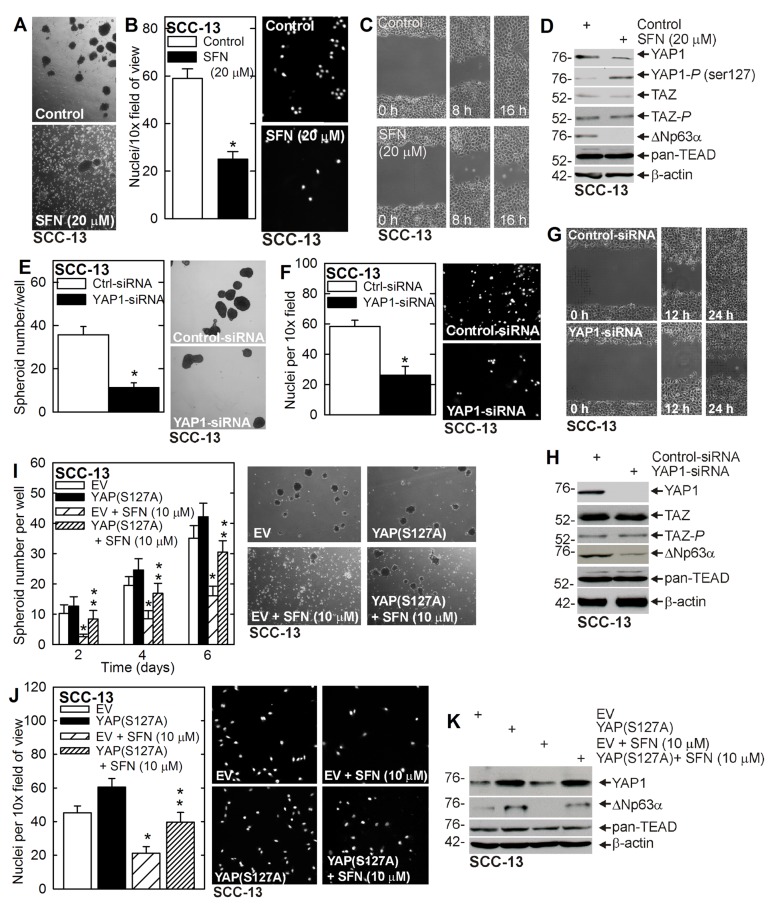
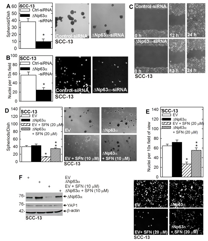
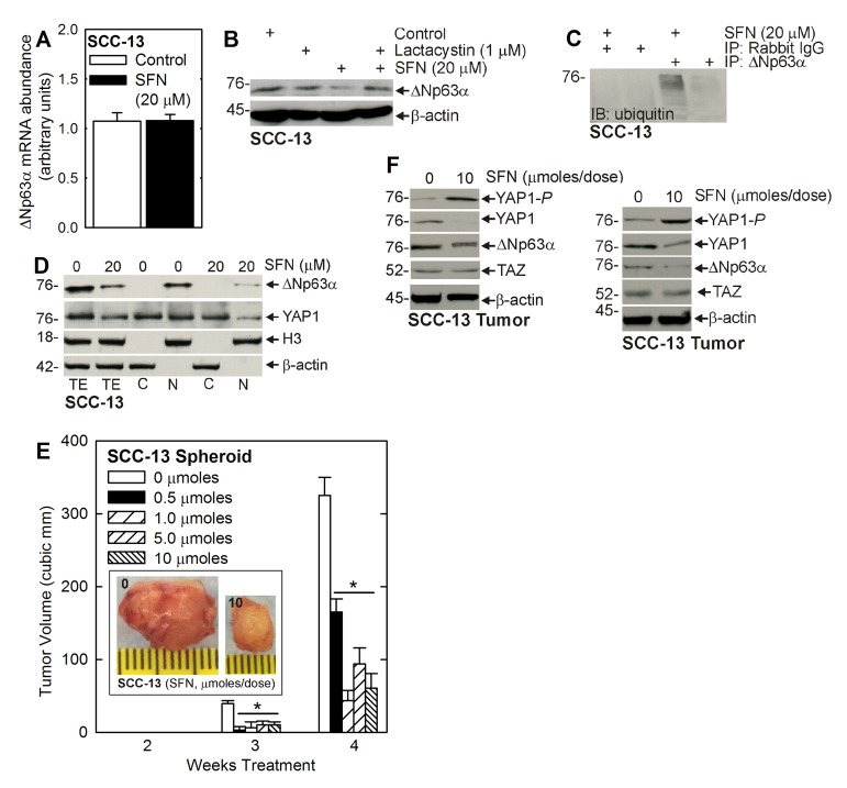
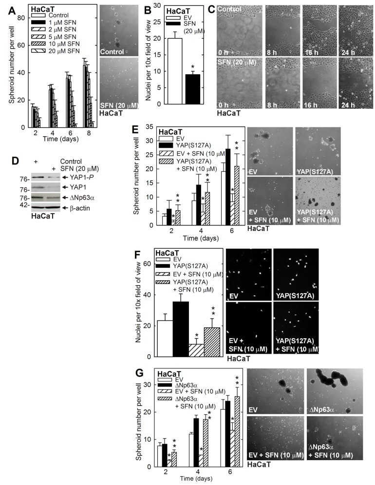
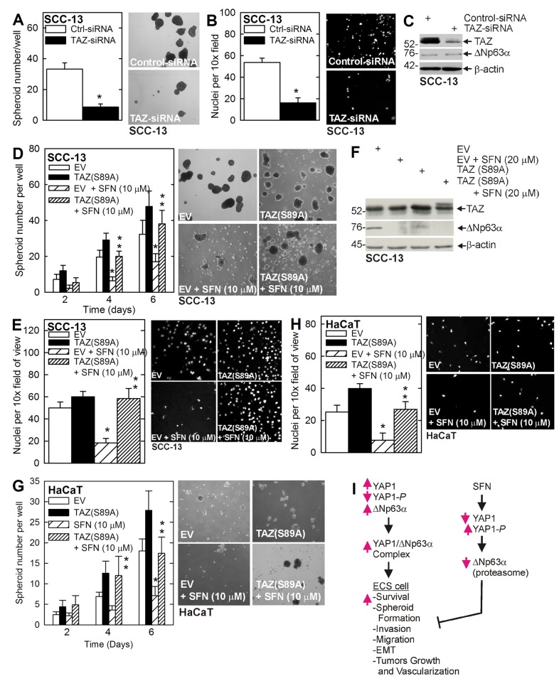
Epidermal squamous cell carcinoma (SCC) is among the most common cancers. SCC can be treated by surgical excision, but recurrence of therapy-resistant disease is a major problem. We recently showed that YAP1, the Hippo signaling transcription adaptor protein, and ∆Np63α, a key epidermal stem cell survival protein, form a complex to drive epidermal cancer stem cell survival. In the present study, we demonstrate that YAP1 and ∆Np63α are important sulforaphane cancer prevention targets. We show that sulforaphane treatment increases YAP1 phosphorylation and proteolytic degradation. The loss of YAP1 is associated with a reduction in ∆Np63α level and a reduction in ECS cell survival, spheroid formation, invasion and migration. Loss of YAP1 and ∆Np63α is mediated by the proteasome and can be inhibited by lactacystin treatment. YAP1 or ∆Np63α knockdown replicates the responses to sulforaphane, and restoration of YAP1 or ∆Np63α antagonizes sulforaphane action. Sulforaphane suppresses ECS cell tumor formation and this is associated with reduced levels of YAP1 and ∆Np63α. These studies suggest that YAP1 and ∆Np63α may be important sulforaphane cancer preventive targets in epidermal squamous cell carcinoma.

Keywords: TAZ; YAP; hippo signaling; sulforaphane; ∆Np63α.


Conflict of interest statement

CONFLICTS OF INTEREST The authors declare no conflict of interest

Figures

Figure 1
Figure 1. Sulforaphane targets YAP1/∆Np63α to suppress ECS cell phenotype
A. B. C. ECS cells were grown for 8 d as spheroids and treated with 0 or 20 μM SFN for 48 h before image acquisition. ECS cells were seeded on a matrigel-coated membrane in a Millicell chamber for invasion assay and then treated with 0 or 20 μM SFN for 20 h. ECS cells were plated as high density confluent monolayers for wound closure assay in the presence of 0 or 20 μM SFN. The values are mean ± SEM and the asterisks indicate a significant reduction (n = 3, p < 0.005). D. SFN treatment reduces YAP1, increases YAP1-P and reduces ∆Np63α. Cells were grown as spheroids for 8 d, treated with 0 or 20 μM SFN for 48 h and lysates were collected for immunoblot. E. F. G. SCC-13 cells were electroporated with control- or YAP1-siRNA and plated for spheroid formation, invasion and migration assay. The values are mean ± SEM and the asterisks indicate a significant reduction (n = 3, p < 0.005). H. YAP1-siRNA treatment reduces YAP1 and ∆Np63α level, but does not impact TAZ or TEAD levels. I. J. SCC-13 cells, electroporated with empty vector (EV) or YAP(S127A), were seeded for spheroid growth or invasion assay in the presence of 0 or 10 μM SFN. Spheroid number was monitored at 6 d. The single asterisk indicates a significant reduction in SFN treated as compared to untreated control cultures. The double asterisks indicate a significant increase as compared to the SFN treated group (n = 3, p < 0.01). K. Immunoblot of extracts prepared from 5 d spheroid cultures (panel I).
Figure 2
Figure 2. ∆Np63α drives the ECS cell phenotype
A. B. C. SCC-13 cells were double electroporated with control- or ∆Np63α-siRNA and seeded for spheroid formation, invasion and migration assay. The values are mean ± SEM and the asterisks indicate a significant reduction (n = 3, p < 0.01). D. E. ECS cells were electroporated with empty vector (EV) or ∆Np63α expression vector, and seeded for spheroid formation and invasion assays in the presence of 0 or 20 μM SFN. The single asterisk indicates a significant reduction in SFN treated as compared to untreated control cultures. The double asterisks indicate a significant increase as compared to the SFN treated group (n = 3, p < 0.01). F. Cells, treated as indicated, were grown in non-attached conditions for 5 days and lysates were prepared for immunoblot.
Figure 3
Figure 3. SFN induced proteasome-dependent loss of ∆Np63α
A. ECS cells were treated with 0 or 20 μM SFN for 48 h and extracts were prepared for assay of ∆Np63α mRNA by qRT-PCR. B. ECS cells were pre-treated with 1 μM lactacystin for 1 h, prior to the addition of 20 μM SFN for 24 h. C. ECS cells were treated with SFN for 48 h and lysates immunoprecipitated with anti-∆Np63α for anti-ubiquitin immunoblot. D. Equal cell equivalents of total (TE), nuclear (N), and cytosolic (C) extract, prepared from control or 48 h SFN treated ECS cells, were electrophoresed for immunoblot detection of ∆Np63α, YAP1, histone 3 (nuclear marker) and β-actin (cytoplasmic marker). E. ECS cells (100,000 cells derived from SCC-13) were injected into each front flank in NSG mice. Beginning at 1 d post-injection, SFN was delivered by gavage, three times per week on alternate days at the indicated number of micromoles/dose. Images represent appearance and size of typical control and SFN-treated 4 wk tumors. The values are mean ± SEM and asterisks indicate significant change compared to control, n = 5 mice (10 tumors), p < 0.01. F Tumors were harvested at 4 wk and extracts were prepared for immunoblot. Blots are shown from two representative tumors.
Figure 4
Figure 4. YAP1 and ∆Np63α and the HaCaT cell response to SFN
A. B. C. HaCaT cells were seeded for spheroid formation, invasion and migration assay. Spheroids were counted and photographed at 8 d. The values are mean ± SEM. A significant reduction was in spheroid number was observed at 4, 6 and 8 d at 10 and 20 μM SFN (n = 3, p < 0.005). D. Cells were grown as spheroids for 8 d and then treated with 20 μM SFN for 48 h prior to collection of lysates for immunoblot. E. F. HaCaT cells were electroporated with empty vector (EV) or YAP(S127A) expression vector and at 24 h post-electroporation were seeded for spheroid formation and invasion assay in the presence of 0 or 20 μM SFN. The image shows 6 d spheroids. G. HaCaT cells were electroporated as indicated and then seeded for spheroid growth assay in the presence of 0 or 10 μM SFN. The images show 6 d spheroids. The values are mean ± SEM. The single asterisk indicates a significant reduction in SFN treated versus untreated control cultures. The double asterisks indicate a significant increase as compared to the SFN treated group (n = 3, p < 0.005).
Figure 5
Figure 5. TAZ stimulates the ECS cell phenotype
A. B. C. ECS cells were electroporated with control- or TAZ-siRNA and then seeded for spheroid formation and 18 h invasion assay. Extracts for immunoblot were prepared at 48 h post-electroporation. The images are 6 d spheroids. The values are mean + SEM, and the asterisk indicates a significant reduction in SFN treated versus untreated control cultures (n = 3, p < 0.005). D. E. F. SCC-13 cells were electroporated as indicated and the cells were seeded for spheroid formation and 18 h invasion assay in the presence of 0 or 10 μM SFN. The images are of 6 d spheroids. The single asterisk indicates a significant reduction in SFN treated versus untreated control cultures. The double asterisks indicate a significant increase as compared to the SFN treated group (n = 3, p < 0.005). Extracts were prepared from 6 d spheroids for immunoblot. G. H. HaCaT cells were electroporated with EV or TAZ(S89A) and then seeded for spheroid and invasion assay with or without 10 μM SFN. The asterisks indicate significance as in panels I. Model of SFN action. YAP1 levels are elevated in ECS cells where it binds to ∆Np63α leading to elevated ∆Np63α levels which drives ECS cell survival, etc. SFN treatment stimulates YAP1 phosphorylation leading to reduced YAP1 level leading to proteasome-dependent loss of ∆Np63α which results in reduced ECS cell survival and reduced tumor growth.

References

    1. Lansbury L, Bath-Hextall F, Perkins W, Stanton W, Leonardi-Bee J. Interventions for non-metastatic squamous cell carcinoma of the skin: systematic review and pooled analysis of observational studies. BMJ. 2013;347:f6153. - PMC - PubMed
    1. Adhikary G, Grun D, Kerr C, Balasubramanian S, Rorke EA, Vemuri M, Boucher S, Bickenbach JR, Hornyak T, Xu W, Fisher ML, Eckert RL. Identification of a population of epidermal squamous cell carcinoma cells with enhanced potential for tumor formation. PLoS One. 2013;8:e84324. - PMC - PubMed
    1. Grun D, Adhikary G, Eckert RL. VEGF-A acts via neuropilin-1 to enhance epidermal cancer stem cell survival and formation of aggressive and highly vascularized tumors. Oncogene. 2016;35:4379–87. - PMC - PubMed
    1. Adhikary G, Grun D, Balasubramanian S, Kerr C, Huang JM, Eckert RL. Survival of skin cancer stem cells requires the Ezh2 polycomb group protein. Carcinogenesis. 2015;36:800–10. - PMC - PubMed
    1. Eckert RL, Fisher ML, Grun D, Adhikary G, Xu W, Kerr C. Transglutaminase is a tumor cell and cancer stem cell survival factor. Mol Carcinog. 2015;54:947–58. - PMC - PubMed