. 2017 Aug 24;8(43):75127-75140.
doi: 10.18632/oncotarget.20561. eCollection 2017 Sep 26.

Stabilization of the transcription factors slug and twist by the deubiquitinase dub3 is a key requirement for tumor metastasis

Affiliations

Stabilization of the transcription factors slug and twist by the deubiquitinase dub3 is a key requirement for tumor metastasis

Yiwei Lin et al. Oncotarget. .

Abstract

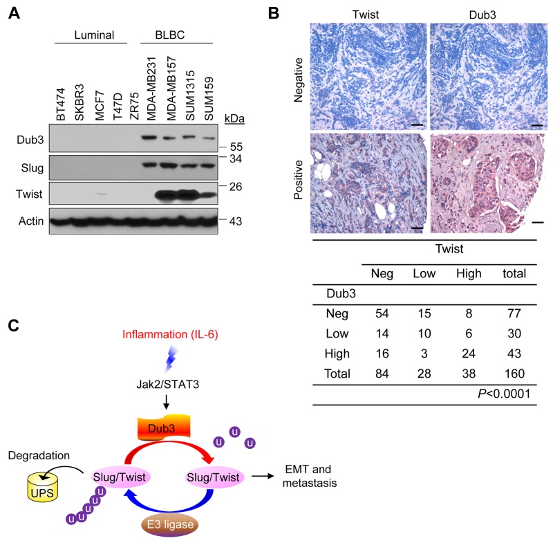
The epithelial-mesenchymal transition (EMT) represents a cellular de-differentiation process that provides cells with the increased plasticity required during embryonic development, tissue remodeling, wound healing and metastasis. Slug and Twist are two key EMT transcription factors (EMT-TFs) that are tightly regulated via ubiquitination and degradation. How Slug and Twist escape degradation and become stabilized in cancer cells remains unclear. One plausible mechanism of Slug and Twist stabilization involves removal of ubiquitin by deubiquitinases (DUBs). In this study, we identified Dub3 as a novel DUB for both Slug and Twist. We further found that Dub3 overexpression increased Slug and Twist protein levels in a dose-dependent manner, whereas Dub3-knockdown decreased their protein levels. Of importance, Dub3 interacted with Slug and Twist and prevented them from degradation, thereby promoting migration, invasion, and cancer stem cell (CSC)-like properties of breast cancer cells. Intriguingly, Dub3 was identified as an early response gene that was upregulated after exposure to inflammatory cytokines such as IL-6, which plays a critical role in the growth and metastasis of breast cancer cells, as well as the maintenance of breast CSCs. We found that Dub3 played an essential role in IL-6 induced EMT through stabilization of Slug and Twist. Our study has uncovered an IL-6-Dub3-Slug/Twist signaling axis during EMT and suggests potential approaches that could target Dub3 to prevent metastatic breast tumor.

Keywords: Dub3; IL-6; Slug; Twist; metastasis.


Conflict of interest statement

CONFLICTS OF INTEREST The authors declare no conflicts of interest.

Figures

Figure 1
Figure 1. Dub3 stabilized Slug and Twist
(A) Flag-tagged Slug and HA-tagged DUBs or HA-Twist and Flag-tagged DUBs were co-expressed in HEK293 cells. After immunoprecipitation, bound Slug or Twist was examined by western blot. (B) Flag-Slug or Flag-Twist was expressed with or without Myc-Dub3 in HEK293 cells or treated with MG132 only. Expression of Slug, Twist and Dub3 was examined by western blot analysis. (C) Flag-Slug or Flag-Twist was co-expressed with wild-type (WT) or C89S (CS) mutant Dub3 in HEK293 cells and examined by western blot analysis. (D) Flag-Slug or Flag-Twist was co-expressed with increasing amounts of Myc-Dub3 in HEK293 cells and examined by western blot analysis. (E) MDA-MB157 and SUM159 cells were stably transfected with control or Dub3 shRNAs and the expression of Slug, Twist and Dub3 was examined by western blot analysis. The mRNAs of Slug, Twist and Dub3 in MDA-MB157 and SUM159 cells stably transfected with control or Dub3 shRNA were examined by real-time PCR. (F) MDA-MB157 cells stably transfected with control or Dub3 shRNA were treated with or without MG132 and expression of Slug, Twist and Dub3 was examined. *p value < 0.001; #p value > 0.05.
Figure 2
Figure 2. Dub3 interacts with Slug and Twist
(A) Flag-Slug or HA-Twist was co-expressed with Myc-Dub3 in HEK293 cells. Slug-Dub3 or Twist-Dub3 complexes were immunoprecipitated with either Myc, HA or Flag antibody, and the associated proteins were examined by western blot analysis using HA, Flag and Myc antibody, respectively. (B) Endogenous Dub3 and Slug or Twist were immunoprecipitated from MDA-MB157 and SUM159 cells, and the bound endogenous Slug/Twist and Dub3 were examined by western blot analysis. (C) EGFP-Slug or EGFP-Twist was co-expressed with Myc-Dub3 in HEK293 cells. After fixation and staining, the cellular location of Slug/Twist (green) and Dub3 (red) was visualized with confocal microscopy. Scale bar = 5 μm.
Figure 3
Figure 3. Dub3 de-ubiquitinates Slug and Twist
(A) Flag-Slug or HA-Twist was co-expressed with Myc-Dub3 or control vector in HEK293 cells. After the cells were treated with cycloheximide (CHX) for indicated time intervals, expression of Slug, Twist and Dub3 was examined by western blot analysis. (B) MDA-MB157 cells stably transfected with control or Dub3 shRNA were treated with CHX. Expression of endogenous Slug, Twist and Dub3 was examined. The intensity of Slug/Twist expression for each time point was quantified by densitometry and plotted (bottom panel). The experiment was repeated three times and a representative experiment is presented. *p value < 0.001; (C) Flag-Slug or Flag-Twist was co-expressed with HA-ubiquitin and either WT or CS Dub3 in HEK293 cells. After cells were treated with MG132, Slug and Twist proteins were immunoprecipitated and poly-ubiquitination of Slug and Twist detected by western blot analysis using HA antibody. (D) MDA-MB157 and SUM159 cells stably transfected with control or Dub3 shRNA were treated with MG132. Cell extracts were immunoprecipitated with Slug or Twist antibody and the poly-ubiquitination of Slug and Twist detected by western blot analysis.
Figure 4
Figure 4. Knockdown of Dub3 inhibits migration, invasion and CSC-like characteristics of BLBC cells
(A) MDA-MB157 and SUM159 cells stably transfected with control or Dub3 shRNA were examined by western blot analysis for expression of various protein markers as indicated. (B) The mRNA levels of various markers as indicated were quantitated by real-time PCR (mean ± SD in three separate experiments). (C) Wound healing assay of MDA-MB157 and SUM159 cells with or without stable knockdown of Dub3. Data were presented as mean ± SEM. (D) Boyden chamber invasion assay of MDA-MB157 and SUM159 cells with or without stable knockdown of Dub3. Data were presented as mean ± SEM. (E) Quantification of tumorsphere from MDA-MB157 and SUM159 cells stably expressing control or Dub3 shRNA (mean ± SD from three independent experiments) (left panel). Representative images were shown (right panel). Scale bar = 100 μm. **p value < 0.01; *p value <0.05.
Figure 5
Figure 5. Overexpression of Dub3 induces EMT
(A) Dub3 was stably transduced in MCF7 and T47D cells and the indicated proteins were examined by western blot analysis. (B) MCF7 and T47D cells with Dub3 overexpression were examined for morphologic changes as well as expression of E-cadherin, ER α and Dub3 with IF. (C) Boyden chamber migration assay of MCF7 and T47D cells with Dub3 WT or CS mutant expression. Data were presented as mean ± SEM. (D) Boyden chamber invasion assay of MCF7 and T47D cells with Dub3 WT or CS mutant expression. Data were presented as mean ± SEM. Scale bar = 100 μm. *p value < 0.001; #p value > 0.05.
Figure 6
Figure 6. IL-6-induced BLBC cells invasion is mediated by Dub3
(A) SUM159 cells were starved overnight and treated with IL-6 for the indicated time intervals and the expression of Dub3, Slug and Twist were examined by western blot analysis. (B) SUM159 cells stably transduced with either control or Dub3 shRNA were starved and treated with IL-6 and the expression of Slug and Twist was examined. (C) SUM159 cells were pre-treated with DMSO, WP1130 or PR619 for half hour. The cells were then treated with IL-6 (50ng/ml) and the expression of Slug and Twist was examined by western blot analysis. (D) SUM159 cells stably transduced with control or Dub3 shRNA were pre-treated with DMSO or WP1130 for half hour. Boyden chamber invasion assay was performed with or without IL-6 induction. Data were presented as mean ± SEM. Scale bar = 100 μm. **p value < 0.01; *p value <0.05; #p value > 0.05.
Figure 7
Figure 7. Dub3, Slug and Twist are coordinately expressed in breast tumors
(A) Expression of Dub3, Slug and Twist was examined in multiple breast cancer cell lines. (B) Breast tumor surgical specimens were immunostained using antibodies against Dub3 and Twist. Representative images of IHC staining from the same tumor samples were shown in the top panel and statistical analysis shown in the bottom panel. (C) The proposed model of the counteracting effect of Dub3 and E3 ligases of Slug and Twist in mediating EMT and metastasis. Scale bar = 100 μm.

References

    1. Nieto MA. The snail superfamily of zinc-finger transcription factors. Nat Rev Mol Cell Biol. 2002;3:155–66. https://doi.org/10.1038/nrm757. - DOI - PubMed
    1. Peinado H, Olmeda D, Cano A. Snail, Zeb and bHLH factors in tumour progression: an alliance against the epithelial phenotype? Nat Rev Cancer. 2007;7:415–28. https://doi.org/10.1038/nrc2131. - DOI - PubMed
    1. Thiery JP, Sleeman JP. Complex networks orchestrate epithelial-mesenchymal transitions. Nat Rev Mol Cell Biol. 2006;7:131–42. https://doi.org/10.1038/nrm1835. - DOI - PubMed
    1. Thiery JP, Acloque H, Huang RY, Nieto MA. Epithelial-mesenchymal transitions in development and disease. Cell. 2009;139:871–90. https://doi.org/10.1016/j.cell.2009.11.007. - DOI - PubMed
    1. Alderton GK. Metastasis: epithelial to mesenchymal and back again. Nat Rev Cancer. 2012;13:3. https://doi.org/10.1038/nrc3428. - DOI - PubMed