

Dissolved molecular hydrogen (H2) in Peritoneal Dialysis (PD) solutions preserves mesothelial cells and peritoneal membrane integrity
- PMID: 29089029
- PMCID: PMC5664574
- DOI: 10.1186/s12882-017-0741-0
Dissolved molecular hydrogen (H2) in Peritoneal Dialysis (PD) solutions preserves mesothelial cells and peritoneal membrane integrity
Abstract
Background: Peritoneal dialysis (PD) is used as renal replacement therapy in patients with end-stage kidney disease. However, peritoneal membrane failure remains problematic and constitutes a critical cause of PD discontinuation. Recent studies have revealed the unique biological action of molecular hydrogen (H2) as an anti-oxidant, which ameliorates tissue injury. In the present study, we aimed to examine the effects of H2 on the peritoneal membrane of experimental PD rats.
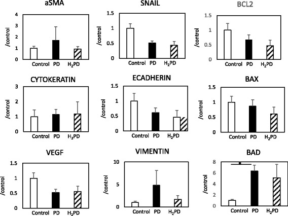
Method: Eight-week-old male Sprague-Dawley rats were divided into the following groups (n = 8-11 each) receiving different test solutions: control group (no treatment), PD group (commercially available lactate-based neutral 2.5% glucose PD solution), and H2PD group (PD solution with dissolved H2 at 400 ppb). Furthermore, the influence of iron (FeCl3: 5 μM: inducer of oxidative cellular injury) in the respective PD solutions was also examined (Fe-PD and Fe-H2PD groups). The H2PD solution was manufactured by bathing a PD bag in H2-oversaturated water created by electrolysis of the water. Twenty mL of the test solutions were intraperitoneally injected once a day for 10 days. Parietal peritoneum samples and cells collected from the peritoneal surface following treatment with trypsin were subjected to analysis.
Results: In the PD group as compared to controls, a mild but significant sub-mesothelial thickening was observed, with increase in the number of cells in the peritoneal surface tissue that were positive for apoptosis, proliferation and vimentin, as seen by immunostaining. There were significantly fewer of such changes in the H2PD group, in which there was a dominant presence of M2 (CD163+) macrophages in the peritoneum. The Fe-PD group showed a significant loss of mesothelial cells with sub-mesothelial thickening, these changes being ameliorated in the Fe-H2PD group.
Conclusion: H2-dissolved PD solutions could preserve mesothelial cells and peritoneal membrane integrity in PD rats. Clinical application of H2 in PD could be a novel strategy for protection of peritoneal tissue during PD treatment.
Keywords: Biocompatibility; Electrolyzed water; Macrophage; Mesothelial cell; Molecular hydrogen; PD solution.
Conflict of interest statement
Consent for publication
Not applicable.
Competing interests
None.
Publisher’s Note
Springer Nature remains neutral with regard to jurisdictional claims in published maps and institutional affiliations.
Figures


References
-
- Popovich RP, Moncrief JW, Nolph KD, Ghods AJ, Twardowski ZJ, Pyle WK. Continuous ambulatory peritoneal dialysis. 1978. J Am Soc Nephrol. 1999;10(4):901–910. - PubMed
-
- Oreopoulos DG, Robson M, Izatt S, Clayton S, deVeber GA. A simple and safe technique for continuous ambulatory peritoneal dialysis (CAPD) Trans Am Soc Artif Intern Organs. 1978;24:484–489. - PubMed
-
- Honda K, Hamada C, Nakayama M, Miyazaki M, Sherif AM, Harada T, Hirano H. Impact of uremia, diabetes, and peritoneal dialysis itself on the pathogenesis of peritoneal sclerosis: a quantitative study of peritoneal membrane morphology. Clin J Am Soc Nephrol. 2008;3(3):720–728. doi: 10.2215/CJN.03630807. - DOI - PMC - PubMed
-
- Kawaguchi Y, Saito A, Kawanishi H, Nakayama M, Miyazaki M, Nakamoto H, Tranaeus A. Recommendations on the management of encapsulating peritoneal sclerosis in Japan, 2005: diagnosis, predictive markers, treatment, and preventive measures. Perit Dial Int. 2005;25(Suppl 4):S83–S95. - PubMed
MeSH terms
Substances
LinkOut - more resources
Full Text Sources
Other Literature Sources
Research Materials
