

Direct observation of the nanoscale Kirkendall effect during galvanic replacement reactions
- PMID: 29089478
- PMCID: PMC5663914
- DOI: 10.1038/s41467-017-01175-2
Direct observation of the nanoscale Kirkendall effect during galvanic replacement reactions
Abstract
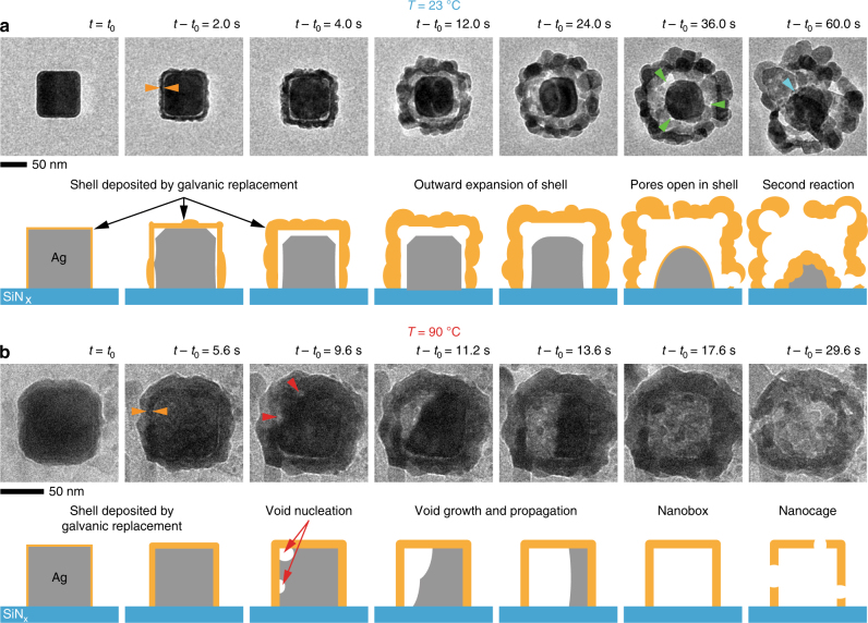
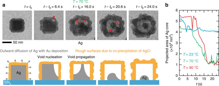
Galvanic replacement (GR) is a simple and widely used approach to synthesize hollow nanostructures for applications in catalysis, plasmonics, and biomedical research. The reaction is driven by the difference in electrochemical potential between two metals in a solution. However, transient stages of this reaction are not fully understood. Here, we show using liquid cell transmission electron microscopy that silver (Ag) nanocubes become hollow via the nucleation, growth, and coalescence of voids inside the nanocubes, as they undergo GR with gold (Au) ions at different temperatures. These direct in situ observations indicate that void formation due to the nanoscale Kirkendall effect occurs in conjunction with GR. Although this mechanism has been suggested before, it has not been verified experimentally until now. These experiments can inform future strategies for deriving such nanostructures by providing insights into the structural transformations as a function of Au ion concentration, oxidation state of Au, and temperature.
Conflict of interest statement
The authors declare no competing financial interests.
Figures


Similar articles
-
Real-Time Dynamics of Galvanic Replacement Reactions of Silver Nanocubes and Au Studied by Liquid-Cell Transmission Electron Microscopy.ACS Nano. 2016 Aug 23;10(8):7689-95. doi: 10.1021/acsnano.6b03020. Epub 2016 Jul 25. ACS Nano. 2016. PMID: 27389989
-
Transformation of Ag nanocubes into Ag-Au hollow nanostructures with enriched Ag contents to improve SERS activity and chemical stability.ACS Appl Mater Interfaces. 2014 Mar 12;6(5):3750-7. doi: 10.1021/am500506j. Epub 2014 Feb 6. ACS Appl Mater Interfaces. 2014. PMID: 24476231
-
In situ liquid cell electron microscopy of Ag-Au galvanic replacement reactions.Nanoscale. 2017 Jan 19;9(3):1271-1278. doi: 10.1039/c6nr07293c. Nanoscale. 2017. PMID: 28054692
-
In Situ Observation of Dynamic Galvanic Replacement Reactions in Twinned Metallic Nanowires by Liquid Cell Transmission Electron Microscopy.Angew Chem Int Ed Engl. 2019 Dec 16;58(51):18627-18633. doi: 10.1002/anie.201910379. Epub 2019 Nov 4. Angew Chem Int Ed Engl. 2019. PMID: 31621994 Review.
-
Interfacially formed organized planar inorganic, polymeric and composite nanostructures.Adv Colloid Interface Sci. 2004 Nov 29;111(1-2):79-116. doi: 10.1016/j.cis.2004.07.005. Adv Colloid Interface Sci. 2004. PMID: 15571664 Review.
Cited by
-
Interior Hotspot Engineering in Ag-Au Bimetallic Nanocomposites by In Situ Galvanic Replacement Reaction for Rapid and Sensitive Surface-Enhanced Raman Spectroscopy Detection.Int J Mol Sci. 2022 Oct 3;23(19):11741. doi: 10.3390/ijms231911741. Int J Mol Sci. 2022. PMID: 36233041 Free PMC article.
-
Butane Dry Reforming Catalyzed by Cobalt Oxide Supported on Ti2 AlC MAX Phase.ChemSusChem. 2020 Dec 7;13(23):6401-6408. doi: 10.1002/cssc.202001633. Epub 2020 Sep 18. ChemSusChem. 2020. PMID: 32945628 Free PMC article.
-
Three-dimensional nanoframes with dual rims as nanoprobes for biosensing.Nat Commun. 2022 Aug 16;13(1):4813. doi: 10.1038/s41467-022-32549-w. Nat Commun. 2022. PMID: 35974015 Free PMC article.
-
Core-shell Au@AuAg nano-peanuts for the catalytic reduction of 4-nitrophenol: critical role of hollow interior and broken shell structure.Nanoscale Adv. 2020 Aug 19;2(10):4841-4852. doi: 10.1039/d0na00312c. eCollection 2020 Oct 13. Nanoscale Adv. 2020. PMID: 36132891 Free PMC article.
-
From Multi- to Single-Hollow Trimetallic Nanocrystals by Ultrafast Heating.Chem Mater. 2023 Nov 6;35(22):9603-9612. doi: 10.1021/acs.chemmater.3c01698. eCollection 2023 Nov 28. Chem Mater. 2023. PMID: 38047181 Free PMC article.
References
-
- Mahmoud MA, O’Neil D, El-Sayed MA. Hollow and solid metallic nanoparticles in sensing and in nanocatalysis. Chem. Mater. 2014;26:44–58. doi: 10.1021/cm4020892. - DOI
Publication types
LinkOut - more resources
Full Text Sources
Other Literature Sources
