. 2017 Oct 31;7(1):14403.
doi: 10.1038/s41598-017-14786-y.

Proton minibeam radiation therapy spares normal rat brain: Long-Term Clinical, Radiological and Histopathological Analysis

Affiliations

Proton minibeam radiation therapy spares normal rat brain: Long-Term Clinical, Radiological and Histopathological Analysis

Yolanda Prezado et al. Sci Rep. .

Abstract

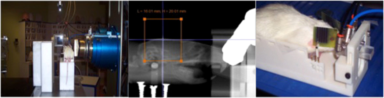
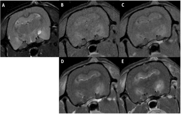
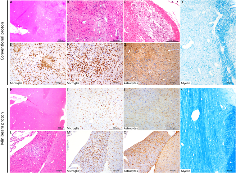
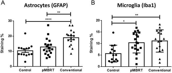
Proton minibeam radiation therapy (pMBRT) is a novel strategy for minimizing normal tissue damage resulting from radiotherapy treatments. This strategy partners the inherent advantages of protons for radiotherapy with the gain in normal tissue preservation observed upon irradiation with narrow, spatially fractionated beams. In this study, whole brains (excluding the olfactory bulb) of Fischer 344 rats (n = 16) were irradiated at the Orsay Proton Therapy Center. Half of the animals received standard proton irradiation, while the other half were irradiated with pMBRT at the same average dose (25 Gy in one fraction). The animals were followed-up for 6 months. A magnetic resonance imaging (MRI) study using a 7-T small-animal MRI scanner was performed along with a histological analysis. Rats treated with conventional proton irradiation exhibited severe moist desquamation, permanent epilation and substantial brain damage. In contrast, rats in the pMBRT group exhibited no skin damage, reversible epilation and significantly reduced brain damage; some brain damage was observed in only one out of the eight irradiated rats. These results demonstrate that pMBRT leads to an increase in normal tissue resistance. This net gain in normal tissue sparing can lead to the efficient treatment of very radio-resistant tumours, which are currently mostly treated palliatively.


Conflict of interest statement

The authors declare that they have no competing interests.

Figures

Figure 1
Figure 1
Experimental setup for rat irradiation (left). Photograph of the Gafchromic films used for quality assurance (right). The orange square represents the irradiation field.
Figure 2
Figure 2
Axial MRI images of one rat in series 1: before Gd injection T2W (A), T1W (B), and T1 FLASH (C), T1W (D) and T1 FLASH after Gd injection (E). Substantial lesions are present. Some hyperintense lesions in the T2W images, along with high signals in the T1W images and BBB breakdown, are compatible with late acute haemorrhage and can be observed in the hippocampus and subiculum. The hypointense lesion in the T2W images of the left hippocampus, along with high signals in the T1W images indicates early subacute haemorrhage. In the right hippocampus, the bright lesion in the T2W images that appears dark in the T1W images and exhibits high intensity in the T1 FLASH images is compatible with late subacute haemorrhage (methaemoglobin). This lesion touches the ventricular system and one part of the brainstem. Extensive BBB breakdown can be observed in the hippocampal formation along with those areas.
Figure 3
Figure 3
Histopathological and immunohistochemical analyses revealed different lesion profiles between the conventional (AF) and minibeam (GL) irradiation groups. A, B, C, H and L: HE staining. D and K: Luxol Fast Blue staining. E, F, I and M: anti-Iba-1 immunohistochemistry (microglia). G, J and O: anti-GFAP immunohistochemistry (astrocytes). The rats were 32 weeks old when sacrificed. Conventional irradiation: (A) Multifocal to coalescing lesion characterized by (B) oedema, necrosis and gliosis. (C) More severe lesion with cavitation and mineralisation. (D) Destruction of the myelin was also observed. (E,F) Microglial activation and microglial nodules (microgliosis). (G) Astrocyte activation with a marked increase in the GFAP immunolabeling (astrogliosis). Minibeam irradiation: (H) At low magnification, no lesion was observed in most rats, with (I,J) normal microglial and astrocytic networks, and (K) normal myelin organization. For just one rat: (L) One inflammatory infiltrate and mild neuropil destruction was observed, associated with focal (M) microgliosis and (O) astrogliosis.
Figure 4
Figure 4
Quantitative morphometric analysis revealed significantly less severe lesions in the pMBRT irradiation group. Activation of astrocytes was significantly stronger in the conventional group (anti-GFAP immunohistochemistry; (A) Besides, both pMBRT and conventional treatment provoked activation of microglial cells (anti-Iba1 immunohistochemistry; (B) *p < 0.05; **p < 0.01; ***p < 0.001; ****p < 0.0001; no star, statistically not significant.

References

    1. Prezado Y, Fois G. Proton-minibeam radiation therapy: A proof of concept. Medical Physics. 2013;40:031712. doi: 10.1118/1.4791648. - DOI - PubMed
    1. Slatkin DN, et al. Subacute neuropathological effects on rats of microplanar beams of x rays from a synchrotron wiggler. Proc. Nat. Acad. Sci. 1995;92:8783–8787. doi: 10.1073/pnas.92.19.8783. - DOI - PMC - PubMed
    1. Dilmanian FA, et al. Interlaced x-ray microplanar beams: A radiosurgery approach with clinical potential. Proc. Natl. Acad. Sci. USA. 2006;103:9709–9714. doi: 10.1073/pnas.0603567103. - DOI - PMC - PubMed
    1. Prezado Y, Renier M, Bravin A. A new method of creating minibeam patterns for synchrotron radiation therapy: a feasibility study. J. Synchr. Radiat. 2009;16:582–586. doi: 10.1107/S0909049509012503. - DOI - PubMed
    1. Dilmanian FA, et al. Response of rat intracranial 9L gliosarcoma to microbeam radiation therapy. Neuro. Oncology. 2002;4:26–38. doi: 10.1215/15228517-4-1-26. - DOI - PMC - PubMed

Publication types

MeSH terms