Review
. 2017 Nov-Dec;18(6):946-956.
doi: 10.3348/kjr.2017.18.6.946. Epub 2017 Sep 21.

Cancer of the Anal Canal: Diagnosis, Staging and Follow-Up with MRI

Affiliations
Review

Cancer of the Anal Canal: Diagnosis, Staging and Follow-Up with MRI

Carole Durot et al. Korean J Radiol. 2017 Nov-Dec.

Abstract

Although a rare disease, anal cancer is increasingly being diagnosed in patients with risk factors, mainly anal infection with the human papilloma virus. Magnetic resonance imaging (MRI) with external phased-array coils is recommended as the imaging modality of choice to grade anal cancers and to evaluate the response assessment after chemoradiotherapy, with a high contrast and good anatomic resolution of the anal canal. MRI provides a performant evaluation of size, extent and signal characteristics of the anal tumor before and after treatment, as well as lymph node involvement and extension to the adjacent organs. MRI is also particularly helpful in the assessment of complications after treatment, and in the diagnosis for relapse of the diseases.

Keywords: Anal cancer; Anus; Diagnosis; Magnetic resonance imaging; Staging.


Figures
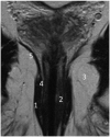
Fig. 1
Fig. 1. Anatomy of anal canal.
T2-weighted coronal MR image. 1 = external sphincter, 2 = internal sphincter, 3 = ischioanal fossa, 4 = puborectal muscle, 5 = levator ani muscle
Fig. 2
Fig. 2. Anatomy of anal canal.
Contrast-enhanced T1-weighted axial image. 1 = external sphincter, 2 = internal sphincter (high enhancement), 3 = fatty intersphincteric space
Fig. 3
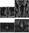
Fig. 3. MR signal of anal canal squamous cell carcinomas.
A, B. Axial (A) and coronal (B) T2-weighted MR images show high signal intensity tumor (arrow) relative to muscles, and of low signal intensity relative to normal ischioanal fat. Lesion is staged T2. C. On low b value of axial diffusion-weighted MR images, lesion shows hyperintensity (arrow). D. Lesion (arrow) is markedly enhanced on fat-suppressed contrast-enhanced T1-weighted axial MR image.
Fig. 4
Fig. 4. Condyloma of anal canal in 53-year-old man.
Sagittal T2-weighted MR image showing condyloma (arrow) displaying cauliflower like appearance.
Fig. 5
Fig. 5. Condyloma with high-grade dysplasia in 45-year-old man with HIV.
Anal margin lesion is seen on axial T2-weighted MR image (arrow, A). Same patient one year later (B, C), axial T2 (B) and fat-suppressed contrast-enhanced T1-weighted (C) MR images show increase in size of lesion (arrow, B) and heterogeneity of enhancement (arrow, C). Histological analysis concluded as squamous cell carcinoma developed on condyloma, T2N0M0.
Fig. 6
Fig. 6. Severe active perineal lesions and multiple fistulas in 50-year-old man with history of Crohn's disease.
Axial fat-suppressed gadolinium-enhanced T1-weighted MR image from first MRI in 2010 (A) shows complex fistula, transphicteric with extension to posterior part of right ischioanal fossa (arrow). Patient was lost to follow-up, and came back six years later. Axial T2 (B) and fat-suppressed gadolinium-enhanced (C) MR weighted images show fistula located at 6 o'clock (arrow, B) as well as large well-circumscribed high signal lesion on T2-weighted MR images (B), not to be misinterpreted as abscess, with inner peripheral enhancement (arrow, C), suggesting mucinous adenocarcinoma developed from long-standing fistula. Histological analysis of resected specimen confirmed this diagnosis.
Fig. 7
Fig. 7. Squamous cell carcinoma of anal canal in 62-year-old man, T2N0M0.
Twenty-two mm lesion is seen invading internal and external sphincter, without extension to adjacent organs, on axial (arrow, A), T2-weighted, high b value (b 1000) (arrow, B), and fat-suppressed gadolnium-enhanced T1-weighted (arrow, C) MR images.
Fig. 8
Fig. 8. Squamous cell carcinoma of anal canal, T4N1aM0, in 73-year-old woman.
Lesion has cranio-caudal extension of 85 mm, invading middle rectum, vagina and uterine cervix. Axial T2 (A), sagittal T2 (B), and axial fat-suppressed contrast-enhanced T1-weighted MR image (C) show extension of lesion to vagina (white arrows, A, B), skip lesion to mid rectum (black arrow, B) and extension to uterine cervix (white arrow, C).
Fig. 9
Fig. 9. Squamous cell carcinoma of anal canal, T4N0M1, in 60-year-old woman.
A. Coronal T2-weighted image shows squamous cell carcinoma of anal canal (arrow). B. Extension of lesion to left piriformis muscle (arrow).
Fig. 10
Fig. 10. Necrotic inguinal lymph node metastasis (arrow).
Fig. 11
Fig. 11. Ano-vaginal fistula in 63-year-old woman, 6 months after chemoradiation therapy for T3N1cM0 squamous cell carcinoma of anal canal.
Axial T2 (A), and fat-suppressed contrast-enhanced T1-weighted MR (B) images display large fistula from treated anterior part of anal canal into vagina (arrows).
Fig. 12
Fig. 12. 63-year-old woman with squamous cell carcinoma of anal canal, T2N1aM0.
Twenty-five-mm lesion of anal canal shows high signal intensity on T2-weighted image (arrow, A), with strong enhancement on contrast-enhanced T1-weighted (arrow, B). MRI control after CRT shows lesion with peripheral enhancement on contrast-enhanced T1-weighted image (arrow, C), corresponding to radionecrosis.

References

    1. American Joint Committee on Cancer. AJCC cancer staging manual. 7th ed. New York: Springer; 2010. pp. 103–106.
    1. Jemal A, Siegel R, Xu J, Ward E. Cancer statistics, 2010. CA Cancer J Clin. 2010;60:277–300. - PubMed
    1. Johnson LG, Madeleine MM, Newcomer LM, Schwartz SM, Daling JR. Anal cancer incidence and survival: the surveillance, epidemiology, and end results experience, 1973-2000. Cancer. 2004;101:281–288. - PubMed
    1. Uronis HE, Bendell JC. Anal cancer: an overview. Oncologist. 2007;12:524–534. - PubMed
    1. Hoots BE, Palefsky JM, Pimenta JM, Smith JS. Human papillomavirus type distribution in anal cancer and anal intraepithelial lesions. Int J Cancer. 2009;124:2375–2383. - PubMed

MeSH terms

Substances