

Sphenoid Sinus Diseases: A Review of 1,442 Patients
- PMID: 29090009
- PMCID: PMC5635283
- DOI: 10.1155/2017/9650910
Sphenoid Sinus Diseases: A Review of 1,442 Patients
Abstract
Objective: To review and report diseases of the sphenoid sinus from the literature and from a university hospital.
Methods: Inpatients' data were retrospectively gathered and reviewed from January 2006 to June 2016. Clinical data, imaging, organisms, and pathological reports were collected. Pathology was divided into infection/inflammation, tumor, and miscellaneous. A literature review was performed with the search term "isolated sphenoid disease" in PubMed. Original primary studies with 20 patients or more were reviewed.
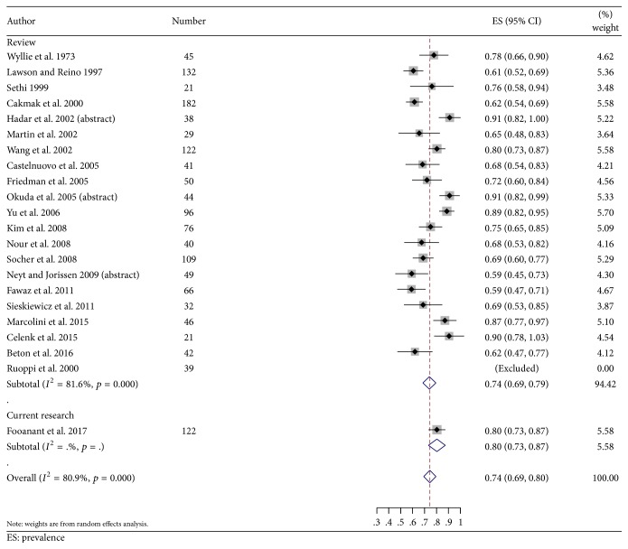
Results and discussion: One hundred and twenty-two patients were enrolled. Seventy-two subjects were female (59%). The average age was 54.3 years (±18.0). Imaging abnormalities were found incidentally in 27 patients (22.1%). The most common symptom was headache (63.9%). Visual loss, the second most common symptom, was more frequent in the tumor group (30.6% versus 54.2%). From the literature review, 21 primary studies with 1,320 total patients were included. From all studies and the present study, infection/inflammation was the most common pathology (75%) [95% confidence interval (CI): 0.696, 0.804]. Overall, tumors were found in 18.9% and malignant tumors in 7.0% [95% CI: 0.045, 0.095].
Conclusion: A specific diagnosis of a sphenoid lesion is needed during active investigation. Infection/inflammation was the most common pathology and malignancy was found in 7%.
Figures


References
Publication types
LinkOut - more resources
Full Text Sources
Other Literature Sources
