Review
. 2017 Oct 25:6:28.
doi: 10.1186/s40035-017-0099-z. eCollection 2017.

Current understanding of the molecular mechanisms in Parkinson's disease: Targets for potential treatments

Affiliations
Review

Current understanding of the molecular mechanisms in Parkinson's disease: Targets for potential treatments

Panchanan Maiti et al. Transl Neurodegener. .

Abstract

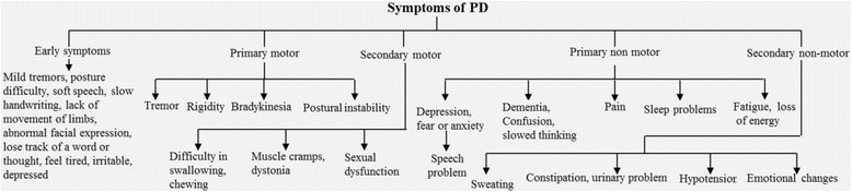
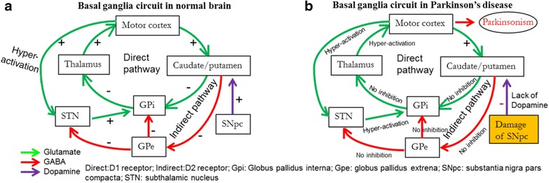
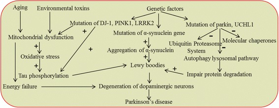
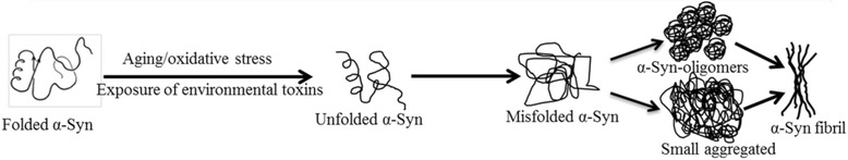
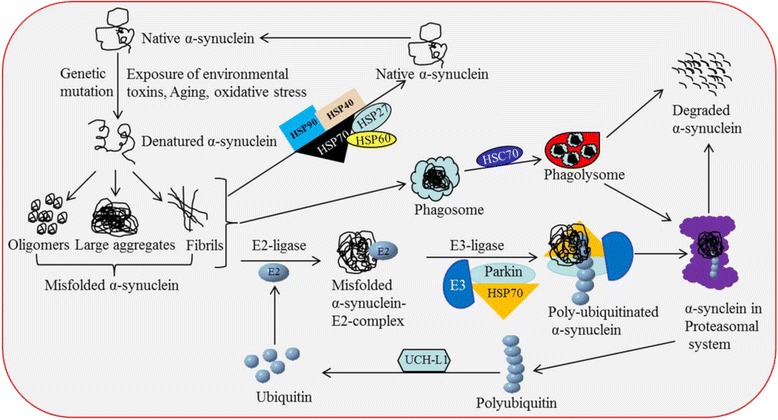
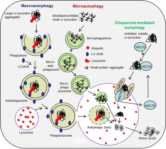
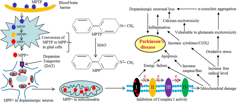
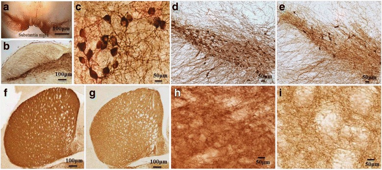
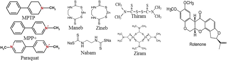
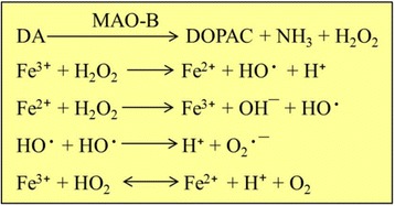
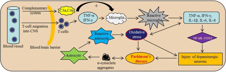
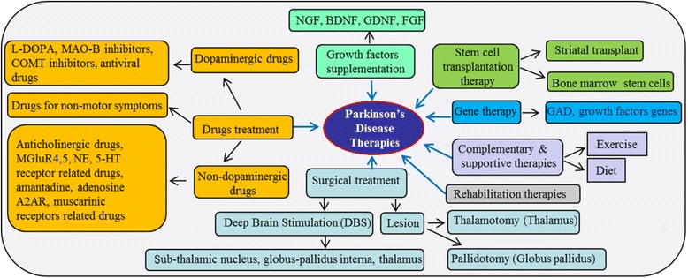
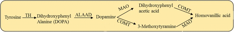
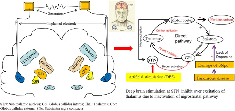
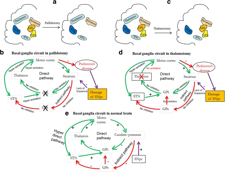
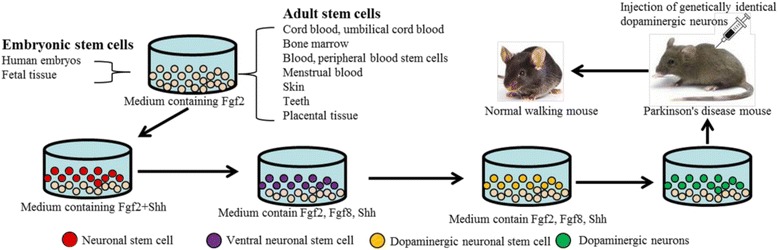
Gradual degeneration and loss of dopaminergic neurons in the substantia nigra, pars compacta and subsequent reduction of dopamine levels in striatum are associated with motor deficits that characterize Parkinson's disease (PD). In addition, half of the PD patients also exhibit frontostriatal-mediated executive dysfunction, including deficits in attention, short-term working memory, speed of mental processing, and impulsivity. The most commonly used treatments for PD are only partially or transiently effective and are available or applicable to a minority of patients. Because, these therapies neither restore the lost or degenerated dopaminergic neurons, nor prevent or delay the disease progression, the need for more effective therapeutics is critical. In this review, we provide a comprehensive overview of the current understanding of the molecular signaling pathways involved in PD, particularly within the context of how genetic and environmental factors contribute to the initiation and progression of this disease. The involvement of molecular chaperones, autophagy-lysosomal pathways, and proteasome systems in PD are also highlighted. In addition, emerging therapies, including pharmacological manipulations, surgical procedures, stem cell transplantation, gene therapy, as well as complementary, supportive and rehabilitation therapies to prevent or delay the progression of this complex disease are reviewed.

Keywords: Cell therapy; Molecular chaperones; Neurodegeneration; Parkinson’s disease; Protein misfolding.


Conflict of interest statement

Ethics approval and consent to participate

Not applicable.

Consent for publication

All authors agreed to publish this article.

Competing interests

The authors declare that they have no competing interests.

Figures

Fig. 1
Fig. 1
Different symptoms of PD. The PD symptoms are categorized into five major subtypes: early, primary motor, secondary motor, primary and secondary non-motor symptoms
Fig. 2
Fig. 2
Neuronal circuits and neurotransmission mechanisms of control in the brains of normal individuals and those with Parkinson’s disease. a: Neuronal circuit in basal ganglia in normal brain. b: Degeneration of substantia nigra pars compacta (SNpc) impairs cortico-striatal circuit in PD brain. Decrease in DA levels in the SNpc and striatum causes loss of control of striatal neuronal firing, leading to withdrawal of inhibitory effects on globus pallidus as well as thalamus, therefore, the thalamus becomes over-excitable, which activates the motor cortex excessively. This ultimately leads to impairment of motor coordination and causes Parkinsonism
Fig. 3
Fig. 3
Schematic diagram showing the involvement of different factors and signaling pathways for degeneration of DA-neurons in PD
Fig. 4
Fig. 4
Schematic diagram showing the steps that cause an accumulation of SNCA. Natural SNCA becomes misfolded under stress and is deposited as oligomers, small aggregates, or fibrils, which play a significant role in DA-neuronal loss in PD
Fig. 5
Fig. 5
Role of protein clearance pathways in PD. Different protein clearance pathways, including molecular chaperones (HSPs), ALP (including macro-autophagy, micro-autophagy and chaperone-mediated autophagy), and the ubiquitin-proteasomal system in degradation of misfolded proteins, such as SNCA and LB have been associated with PD
Fig. 6
Fig. 6
Role of autophagy-lysosomal pathway in degradation of misfolded protein aggregates in PD. Insoluble, larger and smaller SNCA/LB aggregates are degraded by macro-autophagy and micro-autophagy, respectively, whereas soluble, small misfolded SNCA and or LB are degraded by CMA
Fig. 7
Fig. 7
Mechanistic details of MPTP-induced DA-neuronal loss in PD. After crossing blood brain barrier, MPTP enters glial cells, where it is converted to MPP+. This MPP+ then enters neurons and damage mitochondria, which causes energy failure, oxidative stress, glutamate and Ca++ excitotoxicty, increased aggregation of misfolded SNCA, and DA-neuronal loss
Fig. 8
Fig. 8
Brain areas affected by PD. Substantia nigra in mouse brain (a and b); TH+ DA-neurons in SN (c; 40 x); in control (d) and MPTP-treated mouse brain (e). TH+ fibers in control (f, h) and MPTP-treated (g, i) mouse striatum. Note: The loss of DA-neurons in SN (e), along with loss of TH+ fibers in striatum, have been observed after MPTP treatment (g & i)
Fig. 9
Fig. 9
Different ETs-associated with PD. Chemical structure of different pesticides, herbicides, fungicides, and insecticides which may produce Parkinson-like symptoms in animal models
Fig. 10
Fig. 10
Oxidative stress theory in PD. With the help of MAO-B, the DA is converted to DOPAC and produces hydrogen peroxide (H2O2). The H2O2 is then converted to other ROS by Fenton reaction
Fig. 11
Fig. 11
Mechanism of neuroinflammation in PD. T-lymphocytes and complementary systems can activate microglia to secrete several cytokines, which causes DA-neuronal injury. Similarly, aggregated SNCA can also activate astrocytes, which causes oxidative stress, leading to neuronal injury
Fig. 12
Fig. 12
Possible therapies for PD. Currently different therapies available for treating PD include pharmacological manipulations, surgical treatments, stem cell and gene therapies, rehabilitation therapies and other complementary and supportive therapies
Fig. 13
Fig. 13
DA-biosynthesis and degradation. TH: Tyrosine hydroxylase, ALAAD: Aromatic L-amino acid decarboxylase, MAO: Mono amine oxidase, COMT: Catechol O-methyl transferase
Fig. 14
Fig. 14
Schematic diagram show the process of DBS. In DBS, STN or thalamus or the globus pallidus interna (Gpi) (in this case STN) are stimulated by an implanted apparatus contains batteries that produce electrical stimulation (like a pace-maker). Stimulating the STN can activate the GPi, which can strongly inhibit the thalamus (right side circuitry) which can activate the motor cortex; in turn, allowing more control into the movement of limbs
Fig. 15
Fig. 15
Schematic diagram showing pallidotomy (a), and thalamotomy (c) and the basal ganglia circuitory during pallidotomy (b) and thallatomy (d). In case of pallidotomy, the globus pallidus (GP) is surgically destroyed. In the case of a thalamotomy, both thalami are destroyed surgically, which causes a loos of thalamic excitation to the motor cortex, which can decrease Parkinson-like symptoms. Scematic diagram of basal ganglia circuitory in normal brain is shown in “e
Fig. 16
Fig. 16
Different steps of generation of DA-neurons from stem cells for treating PD. Stem cells are obtained from different sources and converted to induced pluoripotent stem cells (iPSCs) using growth factors, such as Fgf2, Shh, Klf4 and c-Myc. The iPSCs is then converted to induced neuronal stem cells (iNSCs). These cells are then converted to DA-neurons by treating different growth factors. These DA-neurons are then injected to the brain of mouse model of PD to supply DA, which ultimately leads to the recovery of motor and cognitive deficits
Fig. 17
Fig. 17
Schematic diagram of basics of rAAV-gene therapy. Left: The gene of interest is packaged within a rAAV vector. When the virus infects the host cell, it injects its DNA-containing gene of interest. This foreign DNA then crosses the nuclear membrane and binds with host DNA. Using protein machinery, the nucleus can make DNA and protein using the inserted DNA, replacing mutated or abnormal genes from host cell. Right: CRISPR-Cas9 system can be used to correct defect gene in PD and other genetic diseases. In presence of guide RNA (g-RNA) CRISPR-Cas9 enzyme can breakdown the DNA double strands in the locus where mutated or faulty genes are located. Then using DNA repair system, the normal DNA can be inserted in the cut site to get normal gene expression

References

    1. Wright Willis A, Evanoff BA, Lian M, Criswell SR, Racette BA. Geographic and ethnic variation in Parkinson disease: a population-based study of US Medicare beneficiaries. Neuroepidemiology. 2010;34(3):143–151. doi: 10.1159/000275491. - DOI - PMC - PubMed
    1. Jankovic J. Parkinson's disease: clinical features and diagnosis. J Neurol Neurosurg Psychiatry. 2008;79(4):368–376. doi: 10.1136/jnnp.2007.131045. - DOI - PubMed
    1. Alexander GE. Biology of Parkinson's disease: pathogenesis and pathophysiology of a multisystem neurodegenerative disorder. Dialogues Clin Neurosci. 2004;6(3):259–280. - PMC - PubMed
    1. Berardelli A, Rothwell JC, Thompson PD, Hallett M. Pathophysiology of bradykinesia in Parkinson's disease. Brain. 2001;124(Pt 11):2131–2146. doi: 10.1093/brain/124.11.2131. - DOI - PubMed
    1. Solari N, Bonito-Oliva A, Fisone G, Brambilla R. Understanding cognitive deficits in Parkinson's disease: lessons from preclinical animal models. Learn Mem. 2013;20(10):592–600. doi: 10.1101/lm.032029.113. - DOI - PubMed