

Nonsyndromic Multiple Basal Cell Carcinomas
- PMID: 29090201
- PMCID: PMC5647844
- DOI: 10.7181/acfs.2017.18.3.191
Nonsyndromic Multiple Basal Cell Carcinomas
Abstract
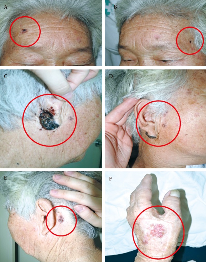
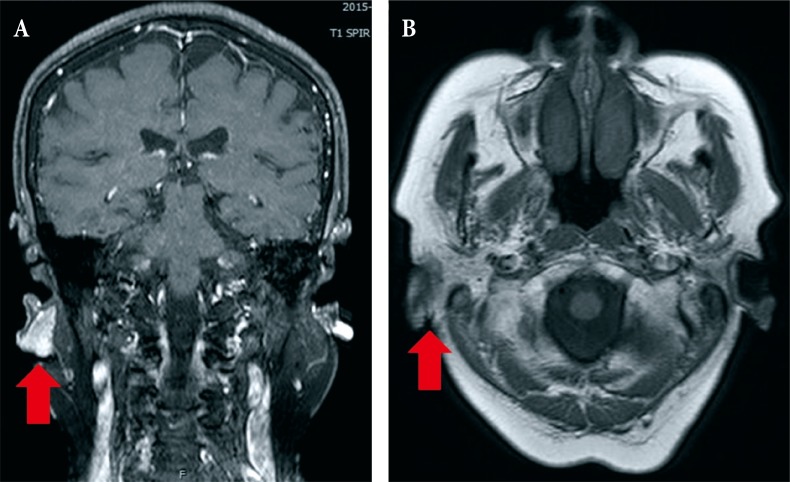
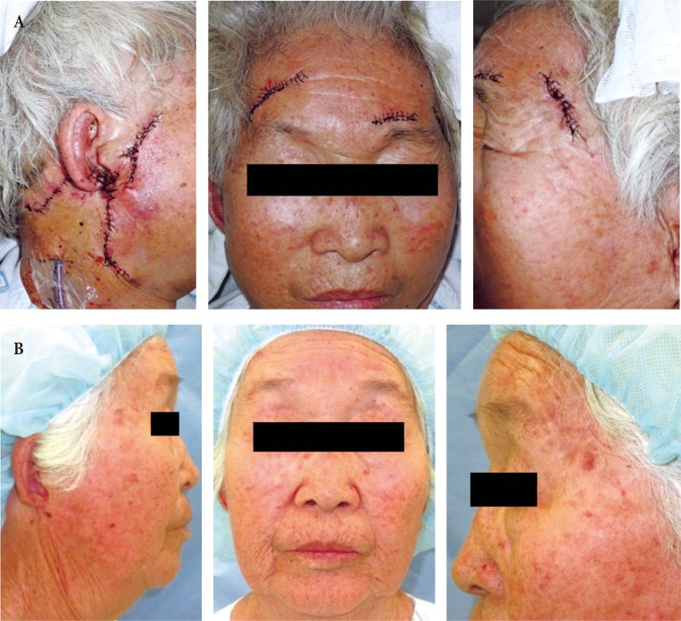
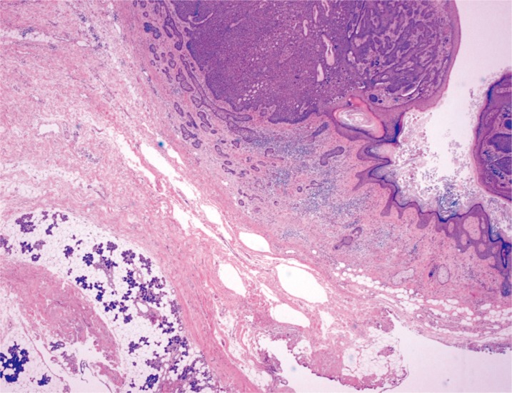
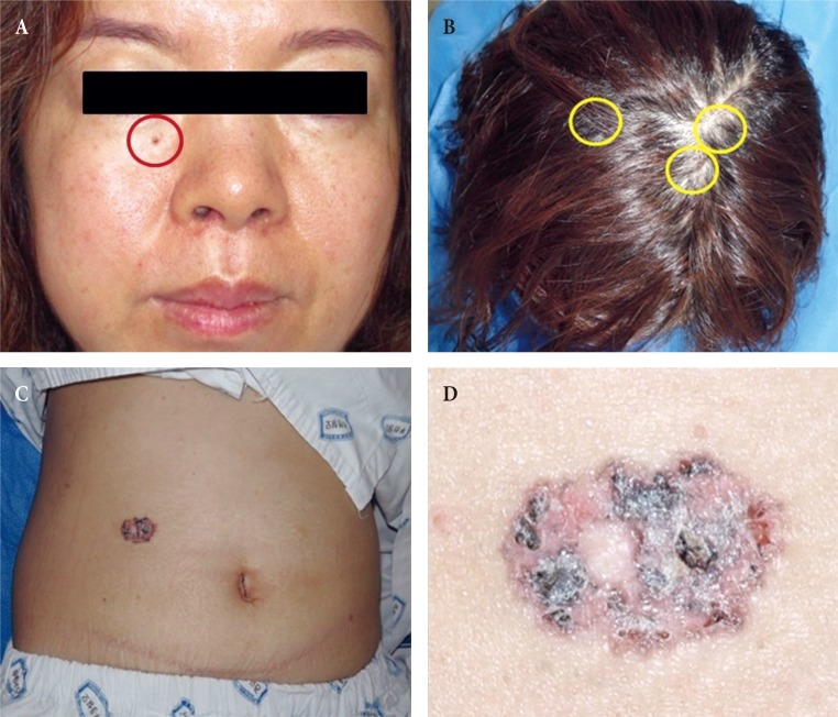
Basal cell carcinoma (BCC) comprising several lesions is not uncommon, but nonsyndromic multiple BCCs with parotid invasion are rare entities. We present two cases of multiple sporadic, nonsyndromic BCCs, and one of these cases is a unique case of parotid invasion associated purely with actinic keratosis. In Case 1, a 79-year-old female presented with multiple skin lesions on the face and left hand. All lesions were completely removed by surgery. The pathologic results showed lesions consistent with BCC and some lesions consistent with actinic keratosis. After 8 months, the patient presented with skin lesions in bilateral temporal areas and left cheek area. Surgical excision of the lesions was performed, and the biopsy results were squamous cell carcinoma in situ and actinic keratosis. In Case 2, a 43-year-old woman presented with multiple skin lesions on the face, scalp, right chest, abdomen and right leg. All lesions were completely removed by surgery. Pathologic evaluation confirmed the diagnosis of BCC. BCC is rarely metastatic, but it can lead to severe disfiguration or destruction. It is important to diagnose and treat BCC at an early stage.
Keywords: Basal cell carcinoma, multiple; Basal cell carcinoma, nonsyndromic; Skin neoplasms.
Conflict of interest statement
No potential conflict of interest relevant to this article was reported.
Figures
References
-
- Zak-Prelich M, Narbutt J, Sysa-Jedrzejowska A. Environmental risk factors predisposing to the development of basal cell carcinoma. Dermatol Surg. 2004;30(2 Pt 2):248–252. - PubMed
-
- McNaughton SA, Marks GC, Green AC. Role of dietary factors in the development of basal cell cancer and squamous cell cancer of the skin. Cancer Epidemiol Biomarkers Prev. 2005;14:1596–1607. - PubMed
-
- Alghamdi Y. Skin tags as a presenting sign of basal cell nevus syndrome in three sisters of the same family. Ann Saudi Med. 2008;28:132–134.
-
- Lu Y, Zhu HG, Ye WM, Zhang MB, He D, Chen WT. A new mutation of PTCH gene in a Chinese family with nevoid basal cell carcinoma syndrome. Chin Med J (Engl) 2008;121:118–121. - PubMed
-
- Farley RL, Manolidis S, Ratner D. Aggressive basal cell carcinoma with invasion of the parotid gland, facial nerve, and temporal bone. Dermatol Surg. 2006;32:307–315. - PubMed
LinkOut - more resources
Full Text Sources
Other Literature Sources
