. 2018 Jul;80(1):317-329.
doi: 10.1002/mrm.26993. Epub 2017 Oct 31.

Relationships between tissue microstructure and the diffusion tensor in simulated skeletal muscle

Affiliations

Relationships between tissue microstructure and the diffusion tensor in simulated skeletal muscle

David B Berry et al. Magn Reson Med. 2018 Jul.

Abstract

Purpose: To establish a series of relationships defining how muscle microstructure and diffusion tensor imaging (DTI) are related.

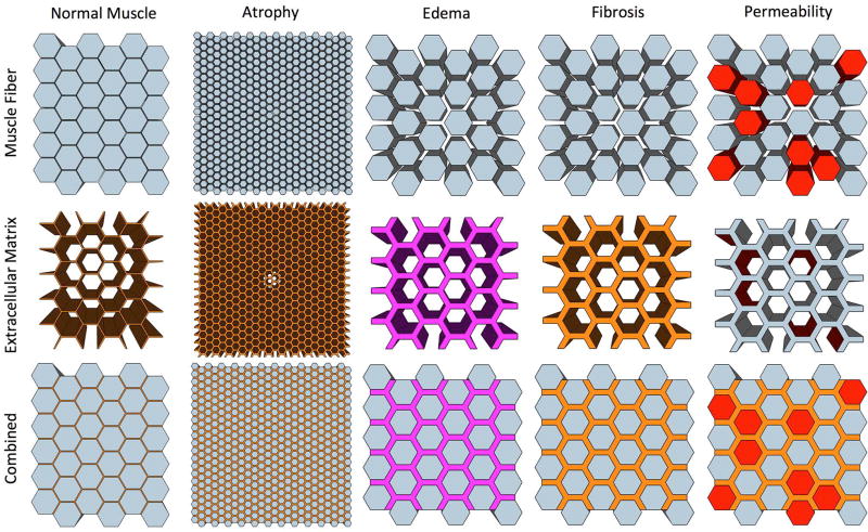
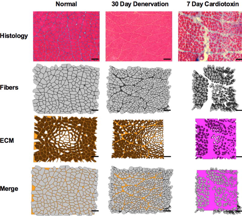
Methods: The relationship among key microstructural features of skeletal muscle (fiber size, fibrosis, edema, and permeability) and the diffusion tensor were systematically simulated over physiologically relevant dimensions individually, and in combination, using a numerical simulation application. Stepwise multiple regression was used to identify which microstructural features of muscle significantly predict the diffusion tensor using single-echo and multi-echo DTI pulse sequences. Simulations were also performed in models with histology-informed geometry to investigate the relationship between fiber size and the diffusion tensor in models with real muscle geometry.

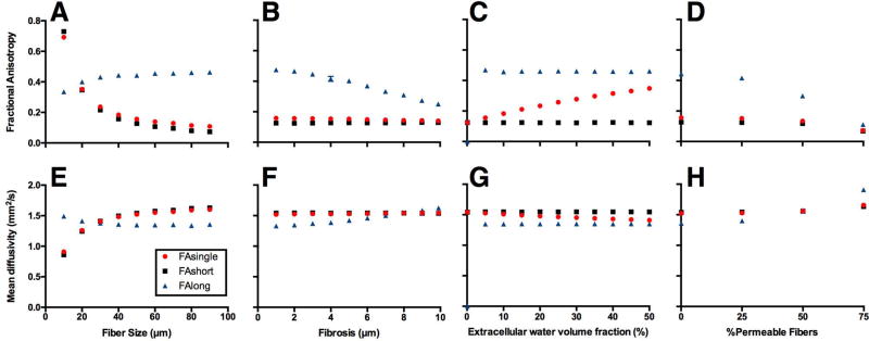
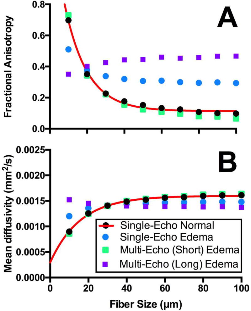
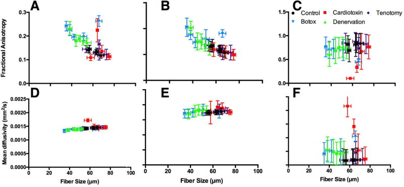
Results: Fiber size is the strongest predictor of λ2, λ3, mean diffusivity, and fractional anisotropy in skeletal muscle, accounting for approximately 40% of the variance in the diffusion model when calculated with single-echo DTI. This increased to approximately 70% when diffusion measures were calculated from the short T2 component of the multi-echo DTI sequence. This nonlinear relationship begins to plateau in fibers with greater than 60-μm diameter.

Conclusions: As the normal fiber size of a human muscle fiber is 40 to 60 μm, this suggests that DTI is a sensitive tool to monitor muscle atrophy, but may be limited in measurements of muscle with larger fibers. Magn Reson Med 80:317-329, 2018. © 2017 International Society for Magnetic Resonance in Medicine.

Keywords: DTI; diffusion; multi-echo DTI; muscle microstructure; simulation; skeletal muscle.


Figures

Figure 1
Figure 1
Schematic depicting ideally shaped hexagonal models of skeletal muscle (top row; intracellular) and the extracellular matrix (middle row; extracellular). Atrophy/hypertrophy was simulated by changing the diameter of the muscle fibers, edema was simulated by changing the extracellular water volume fraction, fibrosis was simulated by changing the spacing between muscle fibers, and permeability was simulated by allowing free transport between fiber walls into the ECM (red).
Figure 2
Figure 2
Schematic depicting sample histology informed models of skeletal muscle. Fibers were manually traced from histology images.
Figure 3
Figure 3
Fractional anisotropy (A–D) and mean diffusivity measurements (E–H) measurements of simplified models of skeletal muscle. Diffusion measurements were made from single-echo DTI (red circles) and the short (black squares) and long (blue triangles) T2 compartments from the multi-echo DTI sequence. Fiber size (A, E), fibrosis (B, F), edema (C, G), and permeability (D, H) were varied over a physiologically relevant range of parameters defined in Table 1.
Figure 4
Figure 4
Fractional anisotropy (A) and mean diffusivity (B) measurements as a function of fiber diameter measured with single-echo (circles) and multi-echo (squares) DTI for normal (black; 5% extracellular water volume fraction) and edematous (45% extracellular water volume fraction) muscle. Non-linear regression (red line) was fit to the normal muscle diffusion measurements measured with single-echo DTI. The equation for the fractional anisotropy regression is FA = (1.432 − 0.1134) * e−0.082*fibersize + 0.1134. The equation for the mean diffusivity regression is MD=2.94104mm2s+(1.30103mm2s)(1e.064fibersize).
Figure 5
Figure 5
Fiber diameter measurements as a function of time for control (black circles), cardiotoxin (red squares), tenotomy (purple diamonds), botox (blue upside down triangles), and denervation (green triangles) models of skeletal muscle. Histology was obtained at day 1, 3, 7, 14, and 30 post-injury from a previous study in our lab.
Figure 6
Figure 6
Fractional anisotropy (A–C) and mean diffusivity (D–F) measurements of models with histology informed geometry from control (black circles), cardiotoxin (red squares), tenotomy (purple diamonds), botox (blue upside down triangles), and denervation (green triangles) skeletal muscle as a function of average muscle fiber diameter. Diffusion measures were made with single-echo (A, D), and the short (B, E) and long (C, F) T2 compartments from the multi-echo DTI sequence.
Figure 7
Figure 7
Fractional anisotropy (A–C) and mean diffusivity (D–F) measurements of models with histology informed geometry from control (black circles), cardiotoxin (red squares), tenotomy (purple diamonds), botox (blue upside down triangles), and denervation (green triangles) skeletal muscle as a function of days after injury. Diffusion measures were made with single-echo (A, D), and the short (B, E) and long (C, F) T2 compartments from the multi-echo DTI sequence.

References

    1. Lieber RL. Skeletal muscle structure, function, and plasticity: Lippincott Williams & Wilkins. 2002
    1. Lieber RL, Ward SR. Cellular mechanisms of tissue fibrosis. 4. Structural and functional consequences of skeletal muscle fibrosis. American Journal of Physiology-Cell Physiology. 2013;305(3):C241–C252. - PMC - PubMed
    1. Minamoto VB, Suzuki KP, Bremner SN, Lieber RL, Ward SR. Dramatic changes in muscle contractile and structural properties after 2 botulinum toxin injections. Muscle & nerve. 2015;52(4):649–657. - PMC - PubMed
    1. Esposito A, Campana L, Palmisano A, De Cobelli F, Canu T, Santarella F, Colantoni C, Monno A, Vezzoli M, Pezzetti G. Magnetic resonance imaging at 7T reveals common events in age-related sarcopenia and in the homeostatic response to muscle sterile injury. PloS one. 2013;8(3):e59308. - PMC - PubMed
    1. Herbison G, Jaweed M, Ditunno J. Muscle fiber atrophy after cast immobilization in the rat. Archives of physical medicine and rehabilitation. 1978;59(7):301–305. - PubMed

Publication types

LinkOut - more resources