. 2018 Mar;11(2):562-574.
doi: 10.1038/mi.2017.74. Epub 2017 Nov 1.

NOX1 loss-of-function genetic variants in patients with inflammatory bowel disease

Collaborators, Affiliations

NOX1 loss-of-function genetic variants in patients with inflammatory bowel disease

T Schwerd et al. Mucosal Immunol. 2018 Mar.

Abstract

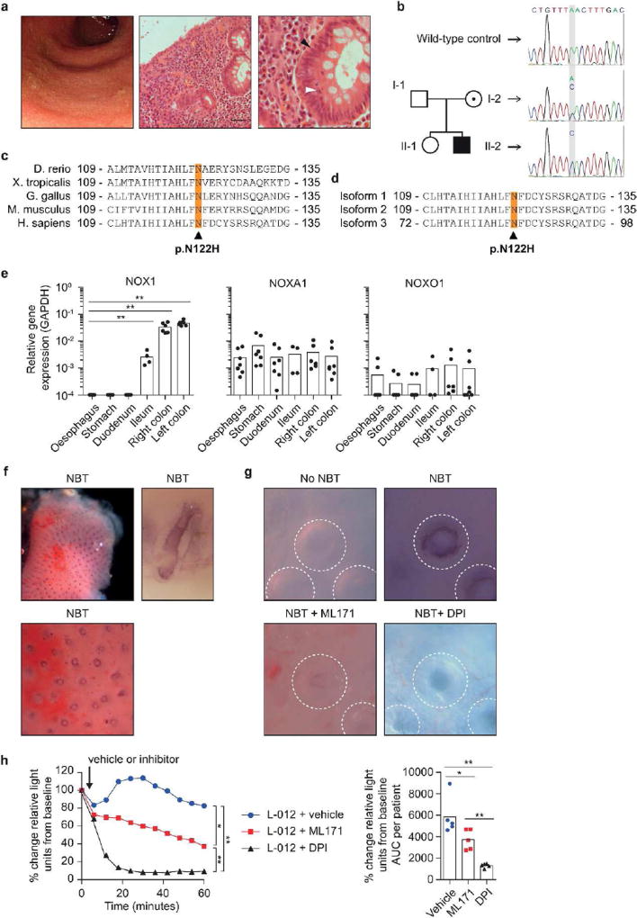
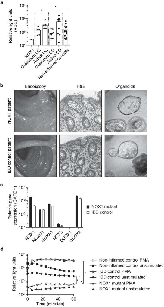
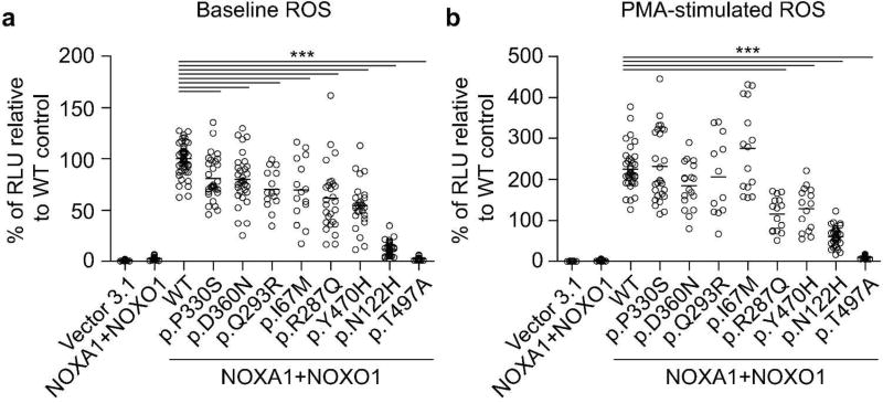
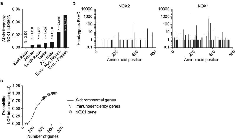
Genetic defects that affect intestinal epithelial barrier function can present with very early-onset inflammatory bowel disease (VEOIBD). Using whole-genome sequencing, a novel hemizygous defect in NOX1 encoding NAPDH oxidase 1 was identified in a patient with ulcerative colitis-like VEOIBD. Exome screening of 1,878 pediatric patients identified further seven male inflammatory bowel disease (IBD) patients with rare NOX1 mutations. Loss-of-function was validated in p.N122H and p.T497A, and to a lesser degree in p.Y470H, p.R287Q, p.I67M, p.Q293R as well as the previously described p.P330S, and the common NOX1 SNP p.D360N (rs34688635) variant. The missense mutation p.N122H abrogated reactive oxygen species (ROS) production in cell lines, ex vivo colonic explants, and patient-derived colonic organoid cultures. Within colonic crypts, NOX1 constitutively generates a high level of ROS in the crypt lumen. Analysis of 9,513 controls and 11,140 IBD patients of non-Jewish European ancestry did not reveal an association between p.D360N and IBD. Our data suggest that loss-of-function variants in NOX1 do not cause a Mendelian disorder of high penetrance but are a context-specific modifier. Our results implicate that variants in NOX1 change brush border ROS within colonic crypts at the interface between the epithelium and luminal microbes.


Conflict of interest statement

Conflict of interest

None of the authors has a conflict of interest related to this article.

Figures

Figure 1
Figure 1. Clinical phenotype of the NOX1 p.N122H patient and NOX1 contribution to baseline ROS within colonic crypts
(a) Endoscopy and histology from NOX1 mutant patient. Colonoscopy shows loss of vascular pattern, ulceration and luminal bleeding (left). H&E stain of colonic biopsies shows crypt architectural distortion and dense inflammatory cell infiltrate in the lamina propria (scale bar 50 µm, middle). Magnification of NOX1 mutant epithelial crypt demonstrates elongated nuclei (black arrow head) and crypt abscesses (white arrow head, right). (b) Family pedigree and Sanger sequencing of NOX1 gene around the mutation site in a wild-type control and family members. (c) NOX1 protein sequence alignment of different genera showing the evolutionary conserved asparagine at position 122. (d) The NOX1 variant p.N122H affects all three isoforms of NOX1. Amino acid sequence was aligned using ClustalW. (e) Analysis of NOX1, NOXA1 and NOXO1 expression by quantitative PCR in biopsies of the gastrointestinal tract. Single biopsies per segment were obtained from 4–7 non-inflamed individuals. Bars represent mean. Statistical significance was determined by Mann-Whitney U test. (f) Ex-vivo colonic biopsies (within 90 minutes of collection) were incubated with nitroblue tetrazolium (NBT) in PBS-Glucose at room temperature. Biopsy specimens were analyzed using light microscopy immediately after 60 minutes NBT incubation (top left, biopsy overview showing dark crypts containing reduced formazan; bottom, ring-like deposition of formazan at epithelial margin of crypts; top right, single colonic epithelial crypt captured longitudinally at high magnification). (g) Microscopy of ROS-producing epithelial cells in colonic crypts of ex-vivo biopsies detected by NBT dye (200× magnification). After 60 minutes of incubation a ring-like deposition of formazan at the epithelial margin of crypts was evident. In indicated conditions biopsies were co-incubated with either a NOX1 inhibitor (ML171) or a non-specific NAPDH oxidase inhibitor (DPI), to verify the signal. (h, left) DPI or ML171 inhibit superoxide generation and L-012-enhanced chemiluminescence on colonic biopsy specimens ex-vivo. Following the detection of superoxide at baseline, DPI, ML171 or vehicle were added (black arrow) and chemiluminescence was recorded over 60 minutes. For each biopsy light emission was normalized to baseline reading. Data represent mean of 9 biopsies per condition obtained from 5 non-inflamed controls. (h, right) Results of kinetic assay calculated as AUC per patient. Statistical significance was determined by Mann-Whitney U test.
Figure 2
Figure 2. NOX1 p.N122H diminishes epithelial ROS in primary organoids
(a) Superoxide generation of colonic epithelial biopsies measured over 60 minutes by L-012-enhanced chemiluminescence and total light emission quantified by area under the curve (AUC). Results are derived from 17 patients with colonic Crohn’s disease (active disease n = 10), 11 patients with ulcerative colitis (active disease n = 6), 16 non-inflamed controls and the patient with NOX1 p.N122H mutation. One symbol represents the mean of 1–3 pan-colonic biopsy specimens (both right and left sided) per patient. Active versus quiescent disease dichotomised according to composite assessment including endoscopic appearance (mucosal ulceration, loss of vascular pattern, bleeding), biomarkers (CRP), and histology (density of inflammatory infiltrate, cryptitis, crypt abscesses). Statistical significance was determined by Mann-Whitney U test. (b) Organoids from patient with NOX1 mutation and IBD control patient were generated from colonic biopsies obtained during quiescent disease state. Representative images of endoscopy, histology (H&E staining, scale bar 50 µm), and organoids are displayed. (c) Analysis of NADPH oxidases and certain subunits by quantitative PCR in NOX1 patient and IBD control organoids. (d) Kinetic assay of PMA-activated L-012-enhanced chemiluminescence on NOX1 mutant, IBD control and non-inflamed control organoid cells. Relative light units (RLU) were measured over 60 minutes. Results are representative of 2 independent experiments measured in replicates. Statistical significance was determined by Wilcoxon signed-rank test comparing the AUC.
Figure 3
Figure 3. Comparative analysis of NOX1 variants
(a, b) Generation of superoxide in colonic epithelial cell line HCT116 reconstituted with multimeric NADPH oxidase complex and expressing wild-type or variant NOX1. ROS was analyzed by L-012-enhanced chemiluminescence in the absence (a) or presence of PMA (b). Chemiluminescence was measured 48 hours post-transfection and recorded over 60 minutes. Results represent eight independent experiments with 3–4 replicates. Each dot represents one replicate. Experiments were normalized to mean relative light units (RLU) without PMA. Line represents mean. Statistical significance was determined with ANOVA and post-test correction performed with Dunnett (multiple comparisons to one single control).
Figure 4
Figure 4. NOX1 population frequencies and genetic variation
(a) Allele frequency of NOX1 p.D360N in different populations according to Exome Aggregation Consortium (ExAC) data and published literature. (b) Number of hemizygotes found for a given variant (missense, nonsense, frameshift) in NOX2 and NOX1 is blotted according to amino acid position (X-axis). Frequency is based on ExAC data. (c) Probability of loss of-function intolerance (pLI) of 727 human X-chromosomal genes based on ExAC data. X-linked primary immunodeficieny genes and NOX1 gene are highlighted.

References

    1. Turner JR. Intestinal mucosal barrier function in health and disease. Nature reviews Immunology. 2009;9(11):799–809. - PubMed
    1. Pastorelli L, De Salvo C, Mercado JR, Vecchi M, Pizarro TT. Central role of the gut epithelial barrier in the pathogenesis of chronic intestinal inflammation: lessons learned from animal models and human genetics. Frontiers in immunology. 2013;4:280. - PMC - PubMed
    1. Khor B, Gardet A, Xavier RJ. Genetics and pathogenesis of inflammatory bowel disease. Nature. 2011;474(7351):307–317. - PMC - PubMed
    1. Jostins L, Ripke S, Weersma RK, Duerr RH, McGovern DP, Hui KY, et al. Host-microbe interactions have shaped the genetic architecture of inflammatory bowel disease. Nature. 2012;491(7422):119–124. - PMC - PubMed
    1. Uhlig HH. Monogenic diseases associated with intestinal inflammation: implications for the understanding of inflammatory bowel disease. Gut. 2013;62(12):1795–1805. - PubMed

Publication types

MeSH terms