. 2018 Jan;285(1):72-86.
doi: 10.1111/febs.14313. Epub 2017 Nov 17.

Fyn-dependent phosphorylation of PlexinA1 and PlexinA2 at conserved tyrosines is essential for zebrafish eye development

Affiliations

Fyn-dependent phosphorylation of PlexinA1 and PlexinA2 at conserved tyrosines is essential for zebrafish eye development

Riley M St Clair et al. FEBS J. 2018 Jan.

Abstract

Plexins (Plxns) are semaphorin (Sema) receptors that play important signaling roles, particularly in the developing nervous system and vasculature. Sema-Plxn signaling regulates cellular processes such as cytoskeletal dynamics, proliferation, and differentiation. However, the receptor-proximal signaling mechanisms driving Sema-Plxn signal transduction are only partially understood. Plxn tyrosine phosphorylation is thought to play an important role in these signaling events as receptor and nonreceptor tyrosine kinases have been shown to interact with Plxn receptors. The Src family kinase Fyn can induce the tyrosine phosphorylation of PlxnA1 and PlxnA2. However, the Fyn-dependent phosphorylation sites on these receptors have not been identified. Here, using mass spectrometry-based approaches, we have identified highly conserved, Fyn-induced PlexinA (PlxnA) tyrosine phosphorylation sites. Mutation of these sites to phenylalanine results in significantly decreased Fyn-dependent PlxnA tyrosine phosphorylation. Furthermore, in contrast to wild-type human PLXNA2 mRNA, mRNA harboring these point mutations cannot rescue eye developmental defects when coinjected with a plxnA2 morpholino in zebrafish embryos. Together these data suggest that Fyn-dependent phosphorylation at two critical tyrosines is a key feature of vertebrate PlxnA1 and PlxnA2 signal transduction.

Keywords: Fyn; mass spectrometry; phosphorylation; plexin; semaphorin.


Figures

Figure 1
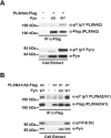
Figure 1. Fyn Induces PLXNA2 and PLXNA1 Phosphorylation
(A–B) HEK293 cells were transfected with expression plasmids encoding PLXNA2-Flag, PLXNA1-Flag, Fyn wildtype (WT) or Fyn kinase dead (KD) as indicated. Cell extracts were subjected to immunoprecipitation with α-Flag resin. Immune complexes and whole cell extracts were subjected to SDS-PAGE and immunoblotting with the indicated antibodies.
Figure 2
Figure 2. Bioinformatic Analysis Identifies a Conserved Tyrosine Phosphorylation Site in PlxnA2 and PlxnA1
(A) PhosphoSitePlus curation shows 20 large-scale mass spectrometry records of human PLXNA2 Y1605 being phosphorylated (top) and 58 records of phosphorylation at the homologous site Y1608 in human PLXNA1 (bottom). Plxns are single-pass type I transmembrane proteins and the N-terminal extracellular and the C-terminal intracellular domain structures are displayed. PSI: plexin, semaphorin, integrin domain; IPT: Ig-like, plexin, transcription factors domain; TM: transmembrane; GAP C1/2: split Ras-GTPase-activating protein domains; RBD: Rho family GTPase-binding domain (B). A sequence alignment highlights the conservation of PlxnA2 Y1605 across vertebrates and invertebrates as well as among PlxnA1 family members. Accession numbers are as follows: human PLXNA2: NP_079455.3, mouse PlxnA2: NP_032908.2, rat PlxnA2: NP_001099458.2, chicken PlxnA2: XP_015154528.1, zebrafish PlxnA2: BAD35133.1, human PLXNA1: NP_115618.3, mouse PlxnA1: NP_032907.1, rat PlxnA1: XP_002729444.2, chicken PlxnA1: XP_414370.4, zebrafish PlxnA1a: XP_003201265.4, zebrafish PlxnA1b: NP_001103480.1, fruit fly PlxnA: NP_524637.2, and roundworm PlxnA: BAB85224.1.
Figure 3
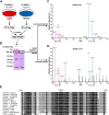
Figure 3. Targeted Mass Spectrometry Reveals Y1605 as a Fyn-Induced Phosphorylation Site on PLXNA2
HEK293 cells were transfected with expression plasmids encoding PLXNA2-Flag, Fyn wildtype (WT) or Fyn kinase dead (KD) as indicated. Whole cell extracts were subjected to immunoprecipitation with α-Flag resin. (A) Whole cell extracts and a small fraction of the immune complexes were subjected to SDS-PAGE and immunoblotting with the indicated antibodies. (B) The majority of the immune complexes for each sample was subjected to SDS-PAGE and protein bands were visualized by coomassie staining. The regions of the gel where PLXNA2 bands run were excised and digested with trypsin and the tryptic peptides were prepared for mass spectrometry. Unphosphorylated and phosphorylated Y1605-containing synthetic reference peptides with a 13C5- and 15N-labeled proline (P*) were added to the native extracted peptides. A targeted approach was used to subject the appropriate m/z values of the doubly-charged tryptic peptides containing either the unphosphorylated or phosphorylated Y1605 to Collision-Induced Dissociation in a linear ion trap-orbitrap mass spectrometer. (C–D) The unphosphorylated native PLXNA2 peptide containing Y1605 was found across treatment groups, but the phosphorylated native peptide was only found in the PLXNA2 + Fyn WT group. Shown are the MS/MS spectra of the phosphorylated Y1605 synthetic peptide (C) and the Y1605 phosphorylated native peptide (D).
Figure 4
Figure 4. Y1677 is a Novel Fyn-Dependent PLXNA2 Phosphorylation Site
(A) Diagram showing the SILAC design to identify PLXNA2 phosphorylation sites dependent on Fyn kinase activity. (B) Coomassie-stained gel of proteins immunoprecipitated from SILAC cell extracts. The region of the gel corresponding to approximately 200 kDa was cut out from experimental and control lanes, subjected to in-gel tryptic digestion and run on a linear ion trap-orbitrap mass spectrometer. (C–D) MS1 m/z windows harboring the isotopic envelopes of the SILAC pairs of tryptic peptide precursor ions of PLXNA2 unphosphorylated at Y1677 (556–564) (C) or phosphorylated at Y1677 596–604 (D). The red peaks represent the isotopic envelopes of the light PLXNA2 peptide precursor ions, or those from the + Fyn WT condition. The blue peaks represent the isotopic envelopes of the heavy PLXNA2 peptide precursor ions, or those from the + Fyn KD condition. The blue, dashed line (bottom) indicates the expected value of the monoisotopic peak of the phosphorylated PLXNA2 precursor peptide ion, which was not present (m/z = 601.282). The black dashed line in both spectra represents the mean heavy:light fold change (mean = 2.87 +/− 0.48 fold change; n = 71, errors bars indicate s.d.) of all other fully tryptic PLXNA2 peptide ions identified. (D) Multiple sequence alignment showing the position of Y1677 across several model organisms for both PlxnA2 as well as PlxnA1.
Figure 5
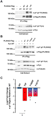
Figure 5. Y1605 and Y1677 are the Major Sites of Fyn-Induced PLXNA2 Tyrosine Phosphorylation
(A–B). HEK293 cells were co-transfected with Fyn WT and Flag-tagged expression constructs for either PLXNA2 WT or the PLXNA2 tyrosine-to-phenylalanine mutants Y2F (Y1605F/Y1677F), Y1605F, or Y1677F. PLXNA2 was immunopurified by α-Flag resin and subjected to SDS-PAGE and immunoblotting with the indicated antibodies. (C) The relative decrease in overall tyrosine phosphorylation normalized to PLXNA2 WT + Fyn WT was quantified for each sample type. Histograms show the average of three independent experiments. *Students T-Test, p = 0.029.
Figure 6
Figure 6. Y1608 and Y1679 are the Major Fyn-Induced Sites of PLXNA1 Tyrosine Phosphorylation
(A). HEK293 cells were co-transfected with Fyn WT or Fyn KD and Flag-tagged expression constructs for either PLXNA1 WT or the PLXNA1 tyrosine-to-phenylalanine Y2F mutant (Y1608F/Y1679F). PLXNA1 was immunopurified by α-Flag resin and subjected to SDS-PAGE and immunoblotting with the indicated antibodies. (B) The relative decrease in overall tyrosine phosphorylation normalized to PLXNA1 WT + Fyn WT was quantified for each sample type. Histograms show the average of three independent experiments. **Students T-Test, WT + Fyn WT comparison to WT + Fyn KD p = 0.005; WT + Fyn WT comparison to Y2F + Fyn WT p = 0.006.
Figure 7
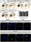
Figure 7. Fyn-dependent PlxnA2 tyrosine phosphorylation sites are essential for zebrafish eye development
(A–F) Brightfield images of zebrafish embryos at 48 hours post fertilization (hpf). (A, B, G) PlxnA2 morphants showed a reduction in eye size compared to uninjected control (eye diameter relative to head length (Control 0.513 µm +/−0.003 µm, n=40 N=4; PlxnA2 MO 0.439 µm +/−0.009 µm, n=40 N=4, ****P<0.0001)). (C, D, G) Co-injection of 2 ng plxna2 MO with 200 pg full-length human PLXNA2-WT mRNA rescued the small eye phenotype, however, full-length human PLXNA2-Y2F mRNA was unable to rescue eye size when co-injected with 2 ng plxna2 MO (Control 0.513 µm +/−0.003 µm, n=40 N=4; WT rescue 0.488 µm +/−0.008 µm, n=34 N=4, n.s; Y2F rescue 0.437 µm +/−0.007 µm, n= 40 N=4, ****P<0.0001). (E, F, G) No observed difference in eye size was seen with overexpression of 200 pg WT or Y2F full-length human mRNA (Control 0.513 µm +/−0.003 µm, n=40 N=4; 200 pg WT 0.493 µm +/−0.005 µm, n=30 N=3, n.s; 200 pg Y2F 0.502 µm +/−0.005 µm, n=30 N=3, n.s). One-way ANOVA, multiple comparisons test. Error bars indicate s.e.m. UIC: uninjected control, MO: morpholino, hpf: hours post fertilization. (H–O) PLXNA2 WT and Y2F show similar subcellular localization in HEK293, COS7, and NIE-115 cells, cell lines that have been used in Sema-Plxn signaling studies. NIE-115 cells show similar PLXNA2 WT and Y2F localization patterns in cells with both long and short processes (L–O). Green is PLXNA2 and blue is DAPI.

References

    1. Maestrini E, Tamagnone L, Longati P, Cremona O, Gulisano M, Bione S, Tamanini F, Neel BG, Toniolo D, Comoglio PM. A family of transmembrane proteins with homology to the MET-hepatocyte growth factor receptor. Proc Natl Acad Sci U S A. 1996;93:674–678. - PMC - PubMed
    1. Jongbloets BC, Pasterkamp RJ. Semaphorin signalling during development. Development. 2014;141:3292–3297. doi: 10.1242/dev.105544. - DOI - PubMed
    1. Kruger RP, Aurandt J, Guan KL. Semaphorins command cells to move. Nat Rev Mol Cell Biol. 2005;6:789–800. doi: 10.1038/nrm1740. - DOI - PubMed
    1. Pasterkamp RJ, Peschon JJ, Spriggs MK, Kolodkin AL. Semaphorin 7A promotes axon outgrowth through integrins and MAPKs. Nature. 2003;424:398–405. doi: 10.1038/nature01790. - DOI - PubMed
    1. Takegahara N, Takamatsu H, Toyofuku T, Tsujimura T, Okuno T, Yukawa K, Mizui M, Yamamoto M, Prasad DV, Suzuki K, et al. Plexin-A1 and its interaction with DAP12 in immune responses and bone homeostasis. Nat Cell Biol. 2006;8:615–622. doi: 10.1038/ncb1416. - DOI - PubMed

Publication types