. 2017 Jan 1;97(6):850-861.
doi: 10.1093/biolre/iox138.

Germline competency of human embryonic stem cells depends on eomesodermin

Affiliations

Germline competency of human embryonic stem cells depends on eomesodermin

Di Chen et al. Biol Reprod. .

Abstract

In humans, germline competency and the specification of primordial germ cells (PGCs) are thought to occur in a restricted developmental window during early embryogenesis. Despite the importance of specifying the appropriate number of PGCs for human reproduction, the molecular mechanisms governing PGC formation remain largely unexplored. Here, we compared PGC-like cell (PGCLC) differentiation from 18 independently derived human embryonic stem cell (hESC) lines, and discovered that the expression of primitive streak genes were positively associated with hESC germline competency. Furthermore, we show that chemical inhibition of TGFβ and WNT signaling, which are required for primitive streak formation and CRISPR/Cas9 deletion of Eomesodermin (EOMES), significantly impacts PGCLC differentiation from hESCs. Taken together, our results suggest that human PGC formation involves signaling and transcriptional programs associated with somatic germ layer induction and expression of EOMES.

Keywords: EOMES; embryonic stem cells; human; primordial germ cells.


Figures

Figure 1.
Figure 1.
Analysis of ITGA6/EPCAM and TNAP/cKIT populations in human prenatal gonads. (A) Flow cytometry of prenatal ovaries at day (d) 72 and 94 postfertilization stained with antibodies that recognize ITGA6, EPCAM, TNAP, and cKIT. (B) Gene expression of the sorted populations from (A, 72d) by real-time PCR. Expression is normalized to the GAPDH. Fold change is calculated relative to expression levels of each gene in female hESC line UCLA1 (passage 17 (p17) and p18), which was given a value of 1.0. (C) Flow cytometry of prenatal testis at day 85 and 104 postfertilization stained with ITGA6, EPCAM, TNAP, and cKIT. (D) Gene expression of the sorted populations from (C, 85d) by real-time PCR. For each gene examined, its expression is normalized to the GAPDH. Fold change is calculated relative to expression levels of each gene in male hESC line UCLA2 (p11 and p12), which was given a value of 1.0. (E) Unsupervised hierarchical clustering (UHC) of transcriptomes of female hESC line UCLA1 (two biological replicates, p14 and p15), male hESC line UCLA2 (two biological replicates, p13 and p14), TNAP/cKIT positive germ cells from embryonic day 59 testes, ITGA6/EPCAM positive cells from 89d, 103d, and another 89d embryonic ovary. TNAP/cKIT positive cells from 89d ovaries. Asterisk in E–G indicates that the two RNA-seq libraries were made from the same pair of ovaries but sorted with different surface markers. (F) PCA of transcriptomes shown in E. (G) Scatter plot of two transcriptomes made from the same pair of ovaries but sorted with ITGA6/EPCAM (x-axis) and TNAP/cKIT (y-axis).
Figure 2.
Figure 2.
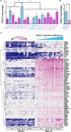
Germline competency varies between independent hESC lines. (A) Average PGCLC induction efficiency at day 4 in aggregates generated from 18 hESC lines (passage numbers ranging from p10 to p22, see experimental procedures for details). Blue represents male and pink represents female. “a*” indicates the significant difference between UCLA6 and all other cell lines and “b*” indicates the significant difference between UCLA2 and UCLA9 (tested by ANOVA, P < 0.0001). PGCLCs were identified as ITGA6/EPCAM double positive cells. (B) Comparison of day 4 PGCLC induction efficiency from male (blue) and female (pink) hESC lines. “*” indicates the difference between male and female (t-test, P < 0.05). (C) Heat map showing the expression of genes in iMeLCs that positively correlated with PGCLC induction efficiency. Genes are selected as maximal expression <= 2 (RPKM) in hESCs, maximal expression >= 2 (RPKM) in iMeLCs, and correlation coefficient >0.45.
Figure 3.
Figure 3.
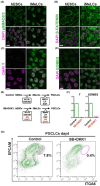
TGFβ and WNT signaling are required for PGCLC induction from hESCs. (A--D) Expression of pSMAD2/3 (A), β-CATENIN (B), T (C), and EOMES (D) in UCLA1 hESCs and iMeLCs. Scale bar: 10 μm. (E) Schematic illustration of PGCLC induction with or without SB431542 (SB) and DKK1. (F) Real-time PCR for T and EOMES expression at iMeLCs in the presence of SB431542 and DKK1. (G) Flow cytometry showing loss of PGCLC competency in media containing SB431542 and DKK1.
Figure 4.
Figure 4.
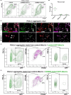
EOMES is required for PGCLC induction from hESCs. (A) Flow cytometry showing reduced PGCLC competency in EOMES mutant hESC line. PGCLCs were identified as ITGA6/EPCAM double positive cells. (B) Summary of PGCLC induction percentage from control and two different EOMES mutant hESC clones. (C) Control and EOMES mutant day 4 aggregates stained with TFAP2C (green), SOX17 (red), PRDM1 (purple), and DAPI (white). White dot outlines TFAP2C/SOX17/PRDM1 triple positive PGCLCs in control. White arrowheads point to rare TFAP2C/SOX17/PRDM1 triple positive PGCLCs in EOMES mutant. (D) Flow cytometry analysis of PGCLCs made from mixed iMeLCs (1:1 ratio) made from GFP negative and GFP positive wild-type hESCs. Left panel shows GFP positive cells in all live cells from PGCLC aggregates. Middle panel shows PGCLCs positive for ITGA6 and EPCAM. Right panel shows GFP positive PGCLCs. (E) Same analysis as (D) for PGCLCs made from mixed iMeLCs (1:1 ratio) made from GFP negative wild-type hESCs and GFP positive EOMES mutant hESCs.

References

    1. Magnusdottir E, Surani MA. How to make a primordial germ cell. Development 2014; 141:245–252. - PubMed
    1. Tang WWC, Kobayashi T, Irie N, Dietmann S, Surani MA. Specification and epigenetic programming of the human germ line. Nat Rev Genet 2016; 17:585–600. - PubMed
    1. Sasaki K, Nakamura T, Okamoto I, Yabuta Y, Iwatani C, Tsuchiya H, Seita Y, Nakamura S, Shiraki N, Takakuwa T, Yamamoto T, Saitou M. The germ cell fate of cynomolgus monkeys is specified in the nascent amnion. Dev Cell 2016; 39:169–185. - PubMed
    1. Clark AT, Gkountela S, Chen D, Liu W, Sosa E, Sukhwani M, Hennebold JD, Orwig KE. Primate primordial germ cells acquire transplantation potential by carnegie stage 23. Stem Cell Reports 2017; 9:329–341. - PMC - PubMed
    1. Kobayashi T, Zhang H, Tang WWC, Irie N, Withey S, Klisch D, Sybirna A, Dietmann S, Contreras DA, Webb R, Allegrucci C, Alberio Ret al. Principles of early human development and germ cell program from conserved model systems. Nature 2017; 546:416–420. - PMC - PubMed

Publication types