. 2017 Dec 22;292(51):20921-20935.
doi: 10.1074/jbc.M117.817023. Epub 2017 Nov 1.

Coordination and redox state-dependent structural changes of the heme-based oxygen sensor Af GcHK associated with intraprotein signal transduction

Affiliations

Coordination and redox state-dependent structural changes of the heme-based oxygen sensor Af GcHK associated with intraprotein signal transduction

Martin Stranava et al. J Biol Chem. .

Abstract

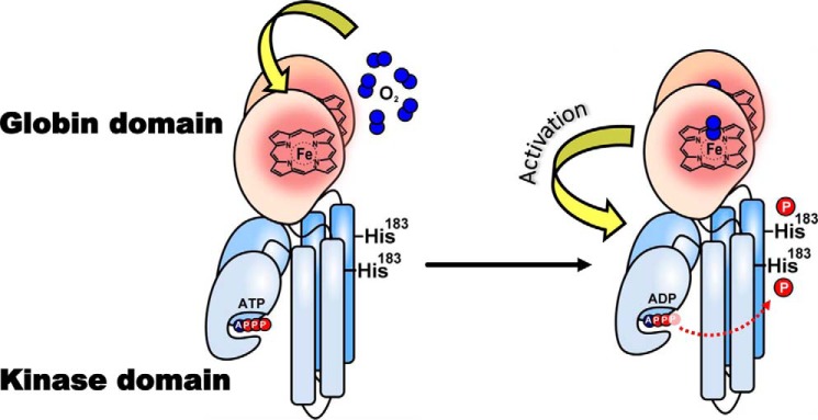
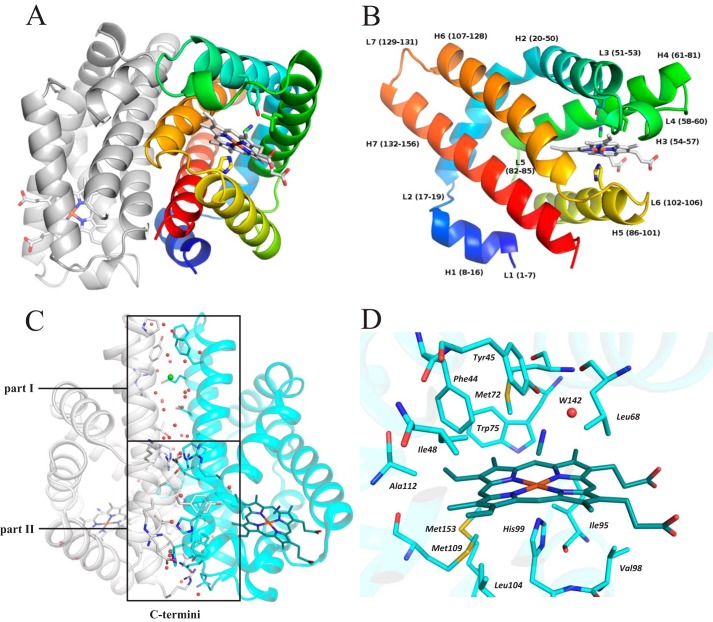
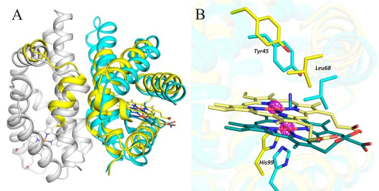
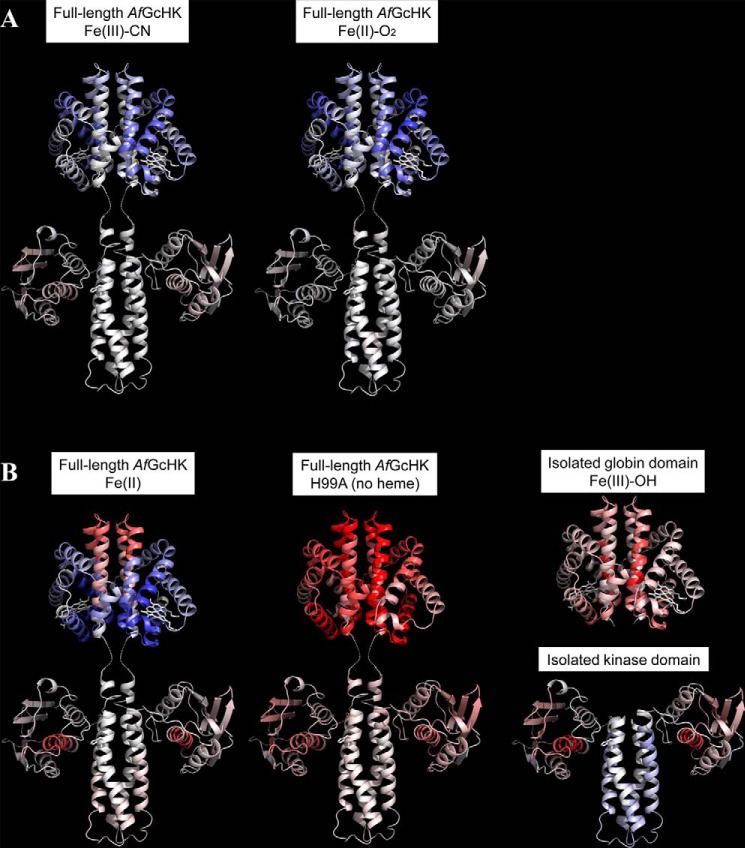
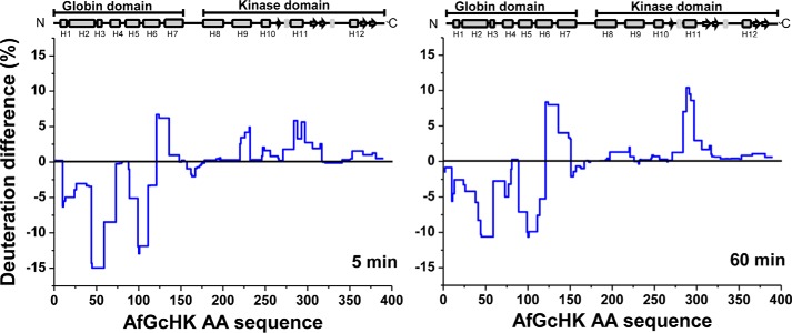
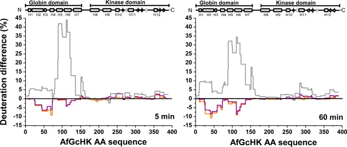
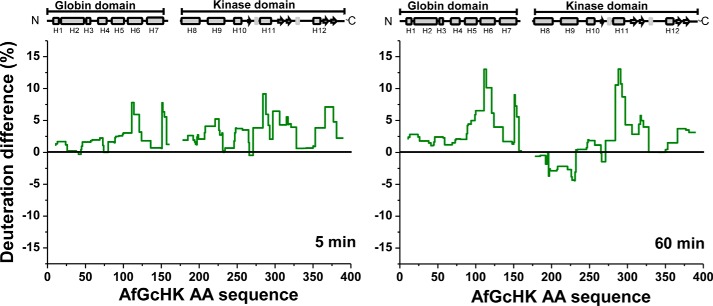
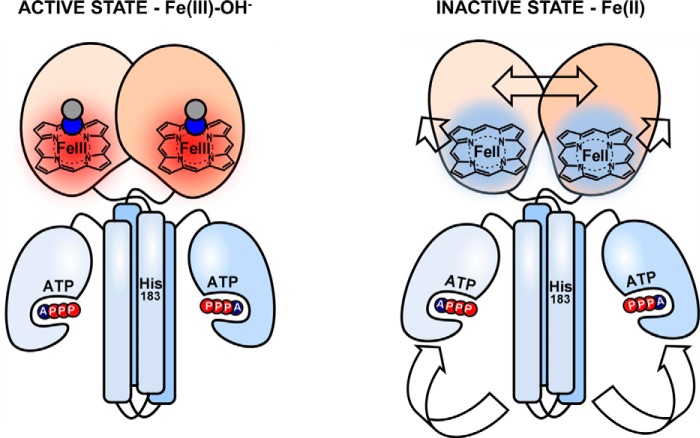
The heme-based oxygen sensor histidine kinase AfGcHK is part of a two-component signal transduction system in bacteria. O2 binding to the Fe(II) heme complex of its N-terminal globin domain strongly stimulates autophosphorylation at His183 in its C-terminal kinase domain. The 6-coordinate heme Fe(III)-OH- and -CN- complexes of AfGcHK are also active, but the 5-coordinate heme Fe(II) complex and the heme-free apo-form are inactive. Here, we determined the crystal structures of the isolated dimeric globin domains of the active Fe(III)-CN- and inactive 5-coordinate Fe(II) forms, revealing striking structural differences on the heme-proximal side of the globin domain. Using hydrogen/deuterium exchange coupled with mass spectrometry to characterize the conformations of the active and inactive forms of full-length AfGcHK in solution, we investigated the intramolecular signal transduction mechanisms. Major differences between the active and inactive forms were observed on the heme-proximal side (helix H5), at the dimerization interface (helices H6 and H7 and loop L7) of the globin domain and in the ATP-binding site (helices H9 and H11) of the kinase domain. Moreover, separation of the sensor and kinase domains, which deactivates catalysis, increased the solvent exposure of the globin domain-dimerization interface (helix H6) as well as the flexibility and solvent exposure of helix H11. Together, these results suggest that structural changes at the heme-proximal side, the globin domain-dimerization interface, and the ATP-binding site are important in the signal transduction mechanism of AfGcHK. We conclude that AfGcHK functions as an ensemble of molecules sampling at least two conformational states.

Keywords: bacterial protein kinase; crystal structure; globin; heme-containing oxygen sensor; histidine kinase; hydrogen-deuterium exchange; signal transduction; two component signal transduction system.


Conflict of interest statement

The authors declare that they have no conflicts of interest with the contents of this article

Figures

Figure 1.
Figure 1.
Intramolecular signal transduction of homodimeric AfGcHK. O2 binds to the heme Fe(II) complex in the globin domain at the N terminus, changing the structure of the protein surrounding the heme. These structural changes function as a signal that is transduced to the kinase domain (at the C terminus), activating the kinase reaction and inducing autophosphorylation at His183.
Figure 2.
Figure 2.
Crystal structure of a dimer of the globin domain of AfGcHK with an Fe(III)-CN heme complex (active state, PDB code 5OHE) and a close-up view of the heme surroundings. A, secondary structure representation of the dimer with the N terminus of chain A in blue, the C terminus of chain A in red, and chain B in gray. The heme, selected neighboring residues, and bound cyanide molecule are shown using a stick representation. B, close-up view of the molecular architecture of protein chain A, using the same color coding and representations as in A; individual α-helices, loops, and the corresponding residue numbers are marked. C, dimerization interface of the isolated globin domain dimer, showing its division into parts I and II; part I is situated opposite the kinase domain in the full-length protein and relies only on van der Waals interactions and water-mediated hydrogen bonds, with no direct protein–protein hydrogen bonds, whereas part II is near the C terminus (close to the kinase domain in the full-length protein) and features 19 interchain protein–protein hydrogen bonds, hydrophobic interactions, and several water-mediated hydrogen bonds. Red dots, water molecules; green sphere, a chloride ion. D, the heme pocket residues (whose carbon atoms are shown in cyan), the heme group (carbon atoms shown in dark cyan), and the cyanide ligand (carbon atom shown in dark cyan) are shown in a stick representation with color coding according to atom type. The graphics were generated using PyMOL (Schrödinger, LLC, New York).
Figure 3.
Figure 3.
Structural changes induced by sodium dithionite soaking (PDB code 5OHF). A, chains G and H are shown using a secondary structure representation (in cyan and gray, respectively) except for their heme and CN moieties, which are shown using a stick representation (colored in dark cyan for chain G and dark gray for chain H). Alternative B is shown in yellow, and the position of its heme group is shown in light yellow using a stick representation. Most of the residues adopting different positions in alternative B are in chain G. B, detailed view of the shift of the heme group and the surrounding heme pocket upon dithionite soaking. The active Fe(III)-CN form is shown in cyan, and the inactive Fe(II) form is shown in yellow. The anomalous difference map (magenta, contoured at 5σ) confirms the presence of iron in two positions (observed in chain G only). The graphics were generated using PyMOL (Schrödinger, LLC).
Figure 4.
Figure 4.
Sedimentation coefficient distributions for the isolated globin domain of AfGcHK (15 μm; black solid line) and the full-length AfGcHK (15 μm; black dashed line). The weight average sedimentation coefficients were calculated from the absorbance data. For further details, see “Experimental procedures.” c(s) denotes the continuous sedimentation coefficient distribution.
Figure 5.
Figure 5.
Conformational changes revealed by HDX–MS after 60 min of deuteration of the full-length AfGcHK proteins and the separated domains, visualized on the protein's structure. Differences between the Fe(III)-OH form and other active or inactive forms are color-coded; gray indicates no difference, whereas higher and lower levels of deuteration in the specified protein form are indicated by increasingly intense red and blue coloration, respectively. A, active full-length AfGcHK proteins with Fe(III)-CN (left) and Fe(II)-O2 (center) heme complexes. B, inactive structures, including the full-length AfGcHK protein with an Fe(II) heme complex (left), the full-length heme-free (apo) H99A mutant (center), the isolated globin domain (top right), and the isolated kinase domain (bottom right). The globin domain structure is based on the X-ray crystal data (PDB code 5OHE) presented in this work, and the kinase domain was modeled as an asymmetric homodimer using subunits A and C of the sensor histidine kinase (HK853) from Thermotoga maritima (PDB code 3DGE). This approach was validated by experiments presented elsewhere (7).
Figure 6.
Figure 6.
HDX–MS protection plots showing the differences in deuteration after 5 min (left) and 60 min (right) of exchange for the full-length Fe(III)-OHbound (active) and Fe(II)-bound (inactive) forms of AfGcHK. The deuteration levels of the active form were subtracted from those of the inactive form and plotted against the protein's sequence (x axis). The blue line thus represents the structural changes caused by reducing the heme iron center in the globin domain of the full-length protein.
Figure 7.
Figure 7.
HDX–MS deuteration differences for the full-length active Fe(II)-O2 (orange) and Fe(III)-CN (purple) forms of AfGcHK and the inactive full-length heme-free H99A mutant (gray) after 5 and 60 min of exchange. The deuteration differences for each form are computed relative to the deuteration of the full-length AfGcHK protein with the Fe(III)-OH heme complex.
Figure 8.
Figure 8.
HDX–MS deuteration differences between the active full-length Fe(III)-OH form of AfGcHK and a mixture of its isolated Fe(III)-OH-bound globin domain with its isolated inactive kinase domain. Deuteration differences are shown for incubation times of 5 and 60 min lined in dark green, and the plotted values were obtained by subtracting the deuteration levels of the active full-length Fe(III)-OH form from those for the isolated domains.
Figure 9.
Figure 9.
Proposed intramolecular signal transduction mechanism in the full-length AfGcHK protein that explains its catalytic inactivation upon heme reduction. Heme reduction induces conformational changes in the globin domain, simultaneously widening and splitting its internal dimeric interface, and making the initial residues of H7 accessible to the solvent. This signal is propagated down to the kinase domain via the linker, causing the ATP binding site to be separated from His183, hindering the autophosphorylation of the latter residue. Data previously obtained by our group indicated that heme reduction increases the enzyme's KmATP value and thus reduces the ATP affinity of the kinase domain (8).

References

    1. Kitanishi K., Kobayashi K., Uchida T., Ishimori K., Igarashi J., and Shimizu T. (2011) Identification and functional and spectral characterization of a globin-coupled histidine kinase from Anaeromyxobacter sp. Fw109-5. J. Biol. Chem. 286, 35522–35534 - PMC - PubMed
    1. Martínková M., Kitanishi K., and Shimizu T. (2013) Heme-based globin-coupled oxygen sensors: linking oxygen binding to functional regulation of diguanylate cyclase, histidine kinase, and methyl-accepting chemotaxis. J. Biol. Chem. 288, 27702–27711 - PMC - PubMed
    1. Gushchin I., Melnikov I., Polovinkin V., Ishchenko A., Yuzhakova A., Buslaev P., Bourenkov G., Grudinin S., Round E., Balandin T., Borshchevskiy V., Willbold D., Leonard G., Büldt G., Popov A., and Gordeliy V. (2017) Mechanism of transmembrane signaling by sensor histidine kinases. Science 10.1126/science.aah6345 - DOI - PubMed
    1. Abriata L. A., Albanesi D., Dal Peraro M., and de Mendoza D. (2017) Signal sensing and transduction by histidine kinases as unveiled through studies on a temperature sensor. Acc. Chem. Res. 50, 1359–1366 - PubMed
    1. Willett J. W., and Crosson S. (2017) Atypical modes of bacterial histidine kinase signaling. Mol. Microbiol. 103, 197–202 - PMC - PubMed

Publication types

LinkOut - more resources