Review
. 2018 Feb;24(2):127-142.
doi: 10.1261/rna.064626.117. Epub 2017 Nov 1.

The RNA exosome and RNA exosome-linked disease

Affiliations
Review

The RNA exosome and RNA exosome-linked disease

Derrick J Morton et al. RNA. 2018 Feb.

Abstract

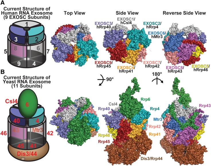
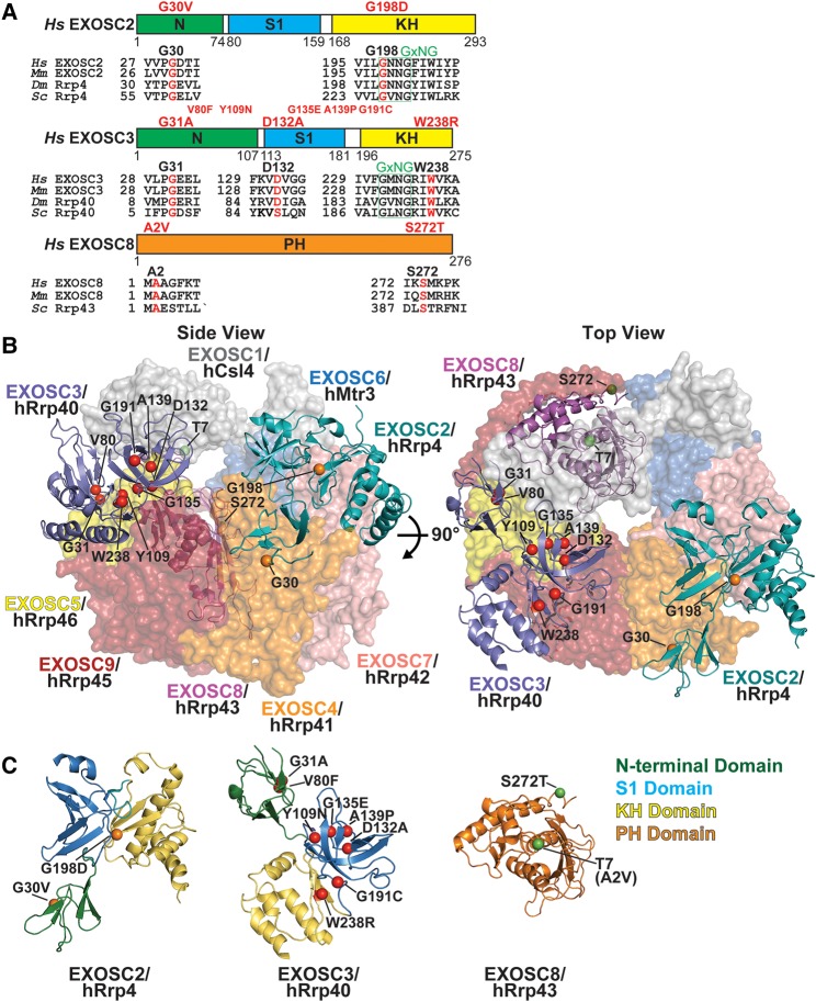
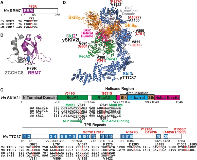
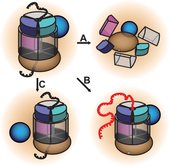
The RNA exosome is an evolutionarily conserved, ribonuclease complex that is critical for both processing and degradation of a variety of RNAs. Cofactors that associate with the RNA exosome likely dictate substrate specificity for this complex. Recently, mutations in genes encoding both structural subunits of the RNA exosome and its cofactors have been linked to human disease. Mutations in the RNA exosome genes EXOSC3 and EXOSC8 cause pontocerebellar hypoplasia type 1b (PCH1b) and type 1c (PCH1c), respectively, which are similar autosomal-recessive, neurodegenerative diseases. Mutations in the RNA exosome gene EXOSC2 cause a distinct syndrome with various tissue-specific phenotypes including retinitis pigmentosa and mild intellectual disability. Mutations in genes that encode RNA exosome cofactors also cause tissue-specific diseases with complex phenotypes. How mutations in these genes give rise to distinct, tissue-specific diseases is not clear. In this review, we discuss the role of the RNA exosome complex and its cofactors in human disease, consider the amino acid changes that have been implicated in disease, and speculate on the mechanisms by which exosome gene mutations could underlie dysfunction and disease.

Keywords: EXOSC2; EXOSC3; EXOSC8; RBM7; RNA exosome; RNA processing/degradation; Rrp4; Rrp40; Rrp43; SKIV2L; Ski2; Ski3; TTC37; intellectual disability; pontocerebellar hypoplasia type 1b; pontocerebellar hypoplasia type 1c; retinitis pigmentosa; spinal motor neuropathy; trichohepatoenteric syndrome.


Figures

FIGURE 1.
FIGURE 1.
Structure of the RNA exosome, a ribonuclease complex that processes/degrades multiple classes of RNA. (A) Cartoon representation of the nine-subunit human RNA exosome complex that has been solved thus far (Liu et al. 2006). This nine-subunit core exosome is composed of a three-subunit cap (EXOSC1/2/3) at the top and a six-subunit ring (EXOSC4-9) in the middle. The active ribonuclease subunit, DIS3, would contact the bottom of this structure, but no human structures have been solved to date that include any DIS3 subunit. To the right of the cartoon, ribbon representations of this nine-subunit human RNA exosome complex (PDB# 2NN6) (Liu et al. 2006) are shown, depicted in top, side, and reverse side views. The nine-subunit human RNA exosome structure reveals a ring-like architecture composed of three cap subunits—EXOSC1/hCsl4 (gray), EXOSC2/hRrp4 (teal), and EXOSC3/hRrp40 (slate blue) and six PH-like ring subunits—EXOSC4/hRrp41 (orange), EXOSC5/hRrp46 (yellow), EXOSC6/hMtr3 (marine blue), EXOSC7/hRrp42 (salmon red), EXOSC8/hRrp43 (magenta), and EXOSC9/hRrp45 (firebrick red). The EXOSC2/hRrp4 (teal) and EXOSC3/hRrp40 (slate blue) cap subunits altered in novel syndrome and pontocerebellar hypoplasia 1b (PCH1b), respectively (Wan et al. 2012; Di Donato et al. 2016), and EXOSC8 (magenta) ring subunit altered in PCH1c (Boczonadi et al. 2014) are highlighted. (B) Cartoon representation of 11-subunit S. cerevisiae RNA exosome, which is composed of a three-subunit cap (Csl4/Rrp4/Rrp40) at the top, a six-subunit ring (Rrp41/Rrp42/Rrp43/Rrp45/Rrp46/Mtr3), and two catalytic subunits (Dis3/Rrp44 and Rrp6) (Makino et al. 2015). To the right of the cartoon, ribbon representations of the structure of this 11-subunit yeast RNA exosome complex (PDB# 5C0W) (Makino et al. 2015) are shown, depicted in top, side, and reverse side views. The 11-subunit yeast RNA exosome structure shows a ring-like shape with three cap subunits—Csl4 (gray), Rrp4 (teal), and Rrp40 (slate blue), six PH-like ring subunits—Rrp41 (orange), Rrp46 (yellow), Mtr3 (marine blue), Rrp42 (salmon red), Rrp43 (magenta), and Rrp45 (firebrick red), and two catalytic subunits—Dis3/Rrp44 (brown) and Rrp6 (forest green). The Rrp6-associated Rrp47 exosome cofactor present in this yeast RNA exosome structure (PDB# 5C0W) (Makino et al. 2015) has been removed. The yeast RNA exosome structure illustrates how the catalytic subunits, Dis3/Rrp44 and Rrp6, interface with the ring-like core of the RNA exosome. The ring-like RNA exosome structures form a central channel through which RNA is directed to the catalytic subunit, Dis3/Rrp44, for processing/degradation. RNA can also gain access to Dis3/Rrp44 in a channel-independent or direct access manner (Liu et al. 2014; Han and van Hoof 2016; Zinder et al. 2016). The color schemes of the human and yeast RNA exosome subunits are identical. Comparison of the human and yeast RNA exosome structures reveals that the RNA exosome structure is highly evolutionarily conserved in eukaryotes.
FIGURE 2.
FIGURE 2.
Amino acid substitutions identified in the EXOSC2, EXOSC3, and EXOSC8 subunits of the RNA exosome in individuals with pontocerebellar hypoplasia and novel syndrome. (A) Domain structures of human EXOSC2, EXOSC3, and EXOSC8 proteins highlighting the amino acid changes identified in affected individuals (Wan et al. 2012; Rudnik-Schöneborn et al. 2013; Zanni et al. 2013; Boczonadi et al. 2014; Eggens et al. 2014; Halevy et al. 2014; Di Donato et al. 2016). Amino acid changes in the EXOSC2 and EXOSC3 cap subunits (shown in red), linked to a novel syndrome and pontocerebellar hypoplasia type 1b (PCH1b), respectively, are located in the N-terminal domain (green), the central putative RNA-binding S1 domain (blue), or the C-terminal putative RNA-binding K homology (KH) domain (yellow). Amino acid changes in the EXOSC8 ring subunit (shown in red), linked to pontocerebellar hypoplasia type 1c (PCH1c), are located in the PH-like domain (orange). Below the domain structures, alignments of EXOSC2/3/8 ortholog sequences from human (Hs), mouse (Mm), Drosophila melanogaster (Dm), and S. cerevisiae (Sc) that surround the evolutionarily conserved residues altered in disease (highlighted in red, labeled in black above) are shown. The GxNG motif (boxed in green) present in the EXOSC2/Rrp4 and EXOSC3/Rrp40 KH domains may play a structural role, as the GXNG motif in ScRrp40 is buried at the interface between the S1 and KH domains (Oddone et al. 2007). Amino acid positions are shown below the domain structures. (B) Structures of the EXOSC2, EXOSC3, and EXOSC8 subunits in the context of the structure of the human RNA exosome complex that highlight the conserved residues altered in disease. The nine-subunit human exosome structure (PDB# 2NN6) (Liu et al. 2006), depicted in top and side views, shows ribbon representations of the EXOSC2/hRrp4 (teal), EXOSC3/hRrp40 (slate blue), and EXOSC8/hRrp43 (magenta) subunits and highlights the conserved residues altered in disease (colored spheres): EXOSC2—G30 and G198 (orange spheres), EXOSC3—G31, V80, Y109, D132, G135, A139, G191, and W238 (red spheres), and EXOSC8—S272 (green sphere), which are labeled in black. The EXOSC8 amino acid T7 (green sphere) is labeled to show the approximate position of the conserved amino acid A2 that is altered in PCH1c individuals, but could not be labeled because it was not resolved in the structure. Transparent, surface representations of the EXOSC1/hCsl4 (gray), EXOSC4/hRrp41 (orange), EXOSC5/hRrp46 (yellow), EXOSC6/hMtr3 (marine blue), EXOSC7/hRrp42 (salmon red), and EXOSC9/hRrp45 (firebrick red) subunits are depicted. (C) Separate ribbon representations of the EXOSC2/hRrp4, EXOSC3/hRrp40, and EXOSC8/hRrp43 subunits that highlight the domains of the proteins and the amino acid substitutions identified in disease. The EXOSC2-G30V and -G198D amino acid substitutions are located in the N-terminal domain (green) and putative RNA-binding KH domain (yellow), respectively. The EXOSC3-G31A and -V80F substitutions are located in the N-terminal domain (green), the EXOSC3-Y109N, -D132A, -G135E, -A139P, and -G191C substitutions are located in the putative RNA-binding S1 domain (blue), and the EXOSC3-W238R substitution is located in the KH domain (yellow). The EXOSC8-S272T substitution is located at the C-terminal end of the PH-like domain (orange). The EXOSC8-T7 residue is labeled to show the approximate position of the EXOSC8-A2V substitution at the N-terminal end of the PH-like domain (orange), as the A2 residue could not be resolved in the structure.
FIGURE 3.
FIGURE 3.
Amino acid substitutions identified in RBM7 of the nuclear exosome cofactor, the NEXT complex, and SKIV2L and TTC37 of the cytoplasmic exosome cofactor, the Ski complex, in individuals with spinal motor neuropathy and syndromic diarrhea/trichohepatoenteric syndrome, respectively. (A) Domain structure of human RBM7 protein highlighting the P79R amino acid change identified in an individual with spinal motor neuropathy (Giunta et al. 2016). The P79R amino acid change in RBM7 (shown in red) is located in the N-terminal RNA recognition motif (RRM) domain (purple). Below the domain structure, alignments of RBM7 ortholog sequences from human (Hs), mouse (Mm), and Drosophila melanogaster (Dm) that surround the evolutionarily conserved residue altered in disease (highlighted in red) are shown. (B) Structure of the RBM7-ZCCHC8 core of the NEXT complex that highlights the conserved P79 residue in RBM7. A ribbon representation of the human RBM7-ZCCHC8 structure (PDB# 5LXR) (Falk et al. 2016) shows the RBM7 RRM (purple) and ZCCHC8 proline-rich region (gray) and highlights the conserved P79 residue altered in disease (dark blue sphere). (C) Domain structures of human SKIV2L and TTC37 proteins highlighting the amino acid changes identified in individuals with trichohepatoenteric syndrome (Hartley et al. 2010; Fabre et al. 2011, 2012; Kammermeier et al. 2014; Oz-Levi et al. 2015; Lee et al. 2016b; Zheng et al. 2016; Kinnear et al. 2017). Amino acid changes in SKIV2L (shown in red), linked to disease, are located in the RecA1 and RecA2 domains (green) in the helicase region. SKIV2L has an N-terminal domain (gray) and C-terminal helicase region, containing two RecA domains (green), a helical bundle domain (pink), and an arch/insertion domain composed of stalk (Stk) regions (brown) and a β-barrel region (light blue). The SKIV2L domain organization is based on sequence alignment with S. cerevisiae Ski2 (Halbach et al. 2012). Amino acid changes in TTC37 (shown in red), linked to disease, are located within and between tetratricopeptide repeat (TPR) motifs (marine blue). TTC37 contains 20 predicted TPR motifs. Below the domain structures, alignments of SKIV2L and TTC37 ortholog sequences from human (Hs), mouse (Mm), Drosophila melanogaster (Dm), and S. cerevisiae (Sc) that surround the evolutionarily conserved residues altered in disease (highlighted in red) are shown. Human SKIV2L and TTC37 residues altered in disease (labeled in black above) and corresponding conserved S. cerevisiae Ski2 and Ski3 residues (labeled in black below) are shown. In SKIV2L, conserved Motif I in RecA1 and Motif IVa in RecA2 (boxed in green) that bind to ATP and nucleic acid, respectively, in other helicases are shown, with specific residues that contact ATP and nucleic acid marked by asterisks (Fairman-Williams et al. 2010; Jankowsky 2011; Johnson and Jackson 2013). (D) Structure of the S. cerevisiae Ski complex, composed of Ski2, Ski3, and two Ski8 proteins, that highlights the positions of conserved residues in yeast Ski2/ySKIV2L and Ski3/yTTC37 altered in SKIV2L and TTC37 in individuals with disease. A ribbon representation of the Ski complex structure (PDB# 4BUJ) (Halbach et al. 2013), depicted in side view, shows Ski2/ySKIV2L (gray; green; pink), Ski3/yTTC37 (marine blue), and two Ski8 proteins Ski8OUT and Ski8IN (located in outer and inner positions in complex) (orange) and highlights the conserved residues altered in disease (red spheres). The human SKIV2L residues—V341 and G631—and TTC37 residues—G673, L761, A1077, and P1270—altered in disease are labeled in red in parentheses and the corresponding, conserved yeast Ski2 residues—V360 and G691—and Ski3 residues—V811, V899, A1155, and V1422 are labeled in black. The Ski2 N-terminal domain (gray), RecA1 and RecA2 domains (green), with Motif I and Motif IVa (yellow), and helicase bundle domain (pink) are depicted.
FIGURE 4.
FIGURE 4.
Potential mechanisms by which disease-linked amino acid changes in EXOSC2, EXOSC3, and EXOSC8 subunits could impair RNA exosome function and lead to tissue-specific phenotypes and diseases. (A) Changes in exosome subunits could disrupt the assembly/disassembly of the RNA exosome complex and impact overall levels of functional complex. (B) Changes in exosome subunits could impair interactions or paths for specific RNA targets (red). (C) Changes in exosome subunits could impair interactions with exosome cofactors (blue sphere). The EXOSC2 (teal), EXOSC3 (slate blue), and EXOSC8 (magenta) exosome subunits are highlighted.

References

    1. Agamy O, Ben Zeev B, Lev D, Marcus B, Fine D, Su D, Narkis G, Ofir R, Hoffmann C, Leshinsky-Silver E, et al. 2010. Mutations disrupting selenocysteine formation cause progressive cerebello-cerebral atrophy. Am J Hum Genet 87: 538–544. - PMC - PubMed
    1. Allmang C, Kufel J, Chanfreau G, Mitchell P, Petfalski E, Tollervey D. 1999a. Functions of the exosome in rRNA, snoRNA and snRNA synthesis. EMBO J 18: 5399–5410. - PMC - PubMed
    1. Allmang C, Petfalski E, Podtelejnikov A, Mann M, Tollervey D, Mitchell P. 1999b. The yeast exosome and human PM-Scl are related complexes of 3′→5′ exonucleases. Genes Dev 13: 2148–2158. - PMC - PubMed
    1. Allmang C, Mitchell P, Petfalski E, Tollervey D. 2000. Degradation of ribosomal RNA precursors by the exosome. Nucleic Acids Res 28: 1684–1691. - PMC - PubMed
    1. Anderson JS, Parker RP. 1998. The 3′ to 5′ degradation of yeast mRNAs is a general mechanism for mRNA turnover that requires the SKI2 DEVH box protein and 3′ to 5′ exonucleases of the exosome complex. EMBO J 17: 1497–1506. - PMC - PubMed

Publication types

MeSH terms

LinkOut - more resources