. 2017 Nov 28;89(22):2288-2296.
doi: 10.1212/WNL.0000000000004690. Epub 2017 Nov 1.

Practice guideline: Cervical and ocular vestibular evoked myogenic potential testing: Report of the Guideline Development, Dissemination, and Implementation Subcommittee of the American Academy of Neurology

Affiliations

Practice guideline: Cervical and ocular vestibular evoked myogenic potential testing: Report of the Guideline Development, Dissemination, and Implementation Subcommittee of the American Academy of Neurology

Terry D Fife et al. Neurology. .

Abstract

Objective: To systematically review the evidence and make recommendations with regard to diagnostic utility of cervical and ocular vestibular evoked myogenic potentials (cVEMP and oVEMP, respectively). Four questions were asked: Does cVEMP accurately identify superior canal dehiscence syndrome (SCDS)? Does oVEMP accurately identify SCDS? For suspected vestibular symptoms, does cVEMP/oVEMP accurately identify vestibular dysfunction related to the saccule/utricle? For vestibular symptoms, does cVEMP/oVEMP accurately and substantively aid diagnosis of any specific vestibular disorder besides SCDS?

Methods: The guideline panel identified and classified relevant published studies (January 1980-December 2016) according to the 2004 American Academy of Neurology process.

Results and recommendations: Level C positive: Clinicians may use cVEMP stimulus threshold values to distinguish SCDS from controls (2 Class III studies) (sensitivity 86%-91%, specificity 90%-96%). Corrected cVEMP amplitude may be used to distinguish SCDS from controls (2 Class III studies) (sensitivity 100%, specificity 93%). Clinicians may use oVEMP amplitude to distinguish SCDS from normal controls (3 Class III studies) (sensitivity 77%-100%, specificity 98%-100%). oVEMP threshold may be used to aid in distinguishing SCDS from controls (3 Class III studies) (sensitivity 70%-100%, specificity 77%-100%). Level U: Evidence is insufficient to determine whether cVEMP and oVEMP can accurately identify vestibular function specifically related to the saccule/utricle, or whether cVEMP or oVEMP is useful in diagnosing vestibular neuritis or Ménière disease. Level C negative: It has not been demonstrated that cVEMP substantively aids in diagnosing benign paroxysmal positional vertigo, or that cVEMP or oVEMP aids in diagnosing/managing vestibular migraine.


Figures

Figure 1
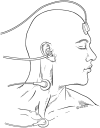
Figure 1. Electrode placement montage for cervical vestibular evoked myogenic potential of the right ear
Surface recording electrodes are placed on the tonically contracted right sternocleidomastoid (achieved by having the patient actively turn the head to the left and lift the head slightly) during recordings. © Barrow Neurological Institute, Phoenix, AZ. Used with permission.
Figure 2
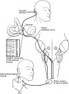
Figure 2. The predominant reflex pathway mediating cervical vestibular evoked myogenic potential responses
Sound stimulates the saccule, which carries the signal through the ipsilateral inferior vestibular nerve to the vestibular nucleus from which vestibulospinal (vestibulocollic) pathway transmits a momentary inhibitory signal to the spinal accessory nerve supplying the ipsilateral sternocleidomastoid muscle. The momentary inhibitory responses are averaged as evoked potentials. I = inferior vestibular nucleus; L = lateral vestibular nucleus; M = medial vestibular nucleus; S = superior vestibular nucleus; VI = abducens nucleus. © Barrow Neurological Institute, Phoenix, AZ. Used with permission.
Figure 3
Figure 3. Cervical vestibular evoked myogenic potential (cVEMP) waveform in a healthy individual
The recordings are from the right sternocleidomastoid muscle in response to 500-Hz tone bursts (2 ms duration, 2 ms rise/fall), with an intensity of 130 dB pSPL. The waveform shows the positive–negative cVEMP (p13-n23) on the side to which the sound stimulation is applied. Measurements include the latency, the amplitude, and the threshold (least intense sound stimulus to still yield a reliable response). The responses occur only on the side of the sound stimulation. Some laboratories display an inverted waveform with the positive wave up instead of the negative wave up. © Barrow Neurological Institute, Phoenix, AZ. Used with permission.
Figure 4
Figure 4. The predominant reflex pathway mediating ocular vestibular evoked myogenic potential responses
Sound stimulates the utricle, which carries the signal through the ipsilateral superior vestibular nerve to the ipsilateral vestibular nucleus. The utriculo-ocular signals cross the midline and travel up the medial longitudinal fasciculus to send an excitatory stimulus to the inferior oblique subnucleus of the oculomotor nucleus, causing momentary contraction which is averaged repetitively. I = inferior vestibular nucleus; III = oculomotor nucleus; L = lateral vestibular nucleus; M = medial vestibular nucleus; MLF = medial longitudinal fasciculus; S = superior vestibular nucleus; VI = abducens nucleus; VN = vestibular nucleus. © Barrow Neurological Institute, Phoenix, AZ. Used with permission.
Figure 5
Figure 5. Ocular vestibular evoked myogenic potential (oVEMP) waveform in a healthy individual
The recordings are from the surface EMG electrodes over the left inferior oblique in response to a sound stimulus in the right ear. The waveform shows the negative–positive oVEMP (n10-p16, sometimes simply referred to as n1 and p1). Measurements include the latency, the amplitude, and the threshold (least intense sound stimulus to still yield a reliable response). The responses occur only on the side opposite the one receiving the sound stimulation. © Barrow Neurological Institute, Phoenix, AZ. Used with permission.

Similar articles

Cited by

References

    1. Colebatch JG, Halmagyi GM. Vestibular evoked potentials in human neck muscles before and after unilateral vestibular differentiation. Neurology 1992;42:1635–1636. - PubMed
    1. Colebatch JG, Halmagyi GM, Skuse NF. Myogenic potentials generated by a click-evoked vestibulocollic reflex. J Neurol Neurosurg Psychiatry 1994;57:190–197. - PMC - PubMed
    1. Clarke AH. Laboratory testing of the vestibular system. Curr Opin Otolaryngol Head Neck Surg 2010;18:425–430. - PubMed
    1. Magliulo G, Gagliardi S, Appiani MC, Iannella G, Re M. Vestibular neurolabyrinthitis: a follow-up study with cervical and ocular vestibular evoked myogenic potentials and the video head impulse test. Ann Otol Rhinol Laryngol 2014;123:162–173. - PubMed
    1. Murofushi T. Clinical application of vestibular evoked myogenic potentials (VEMP). Auris Nasus Larynx 2016;43:367–376. - PubMed

MeSH terms