. 2018 Jan 2;92(2):e01257-17.
doi: 10.1128/JVI.01257-17. Print 2018 Jan 15.

A 3.0-Angstrom Resolution Cryo-Electron Microscopy Structure and Antigenic Sites of Coxsackievirus A6-Like Particles

Affiliations

A 3.0-Angstrom Resolution Cryo-Electron Microscopy Structure and Antigenic Sites of Coxsackievirus A6-Like Particles

Jinhuan Chen et al. J Virol. .

Abstract

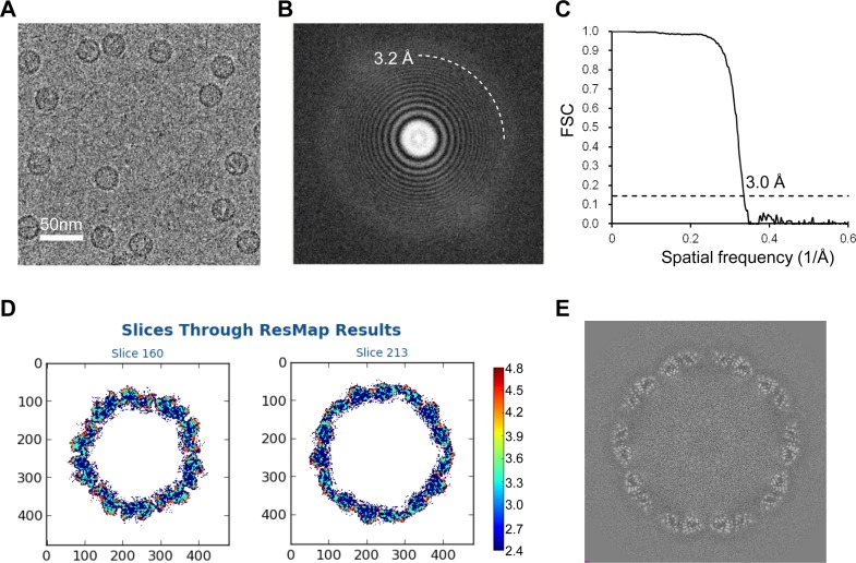
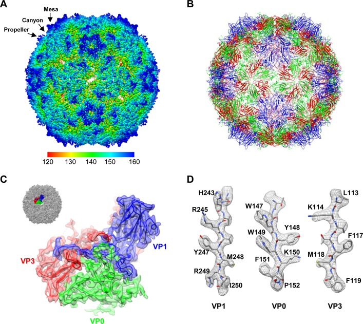
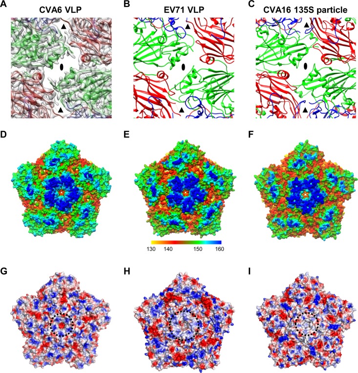
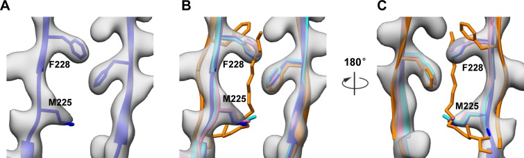
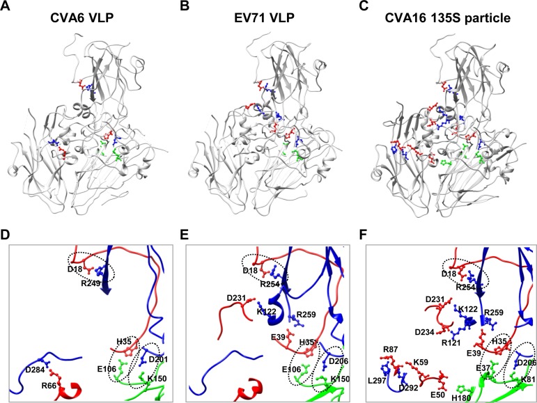
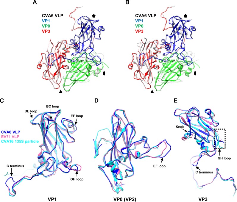
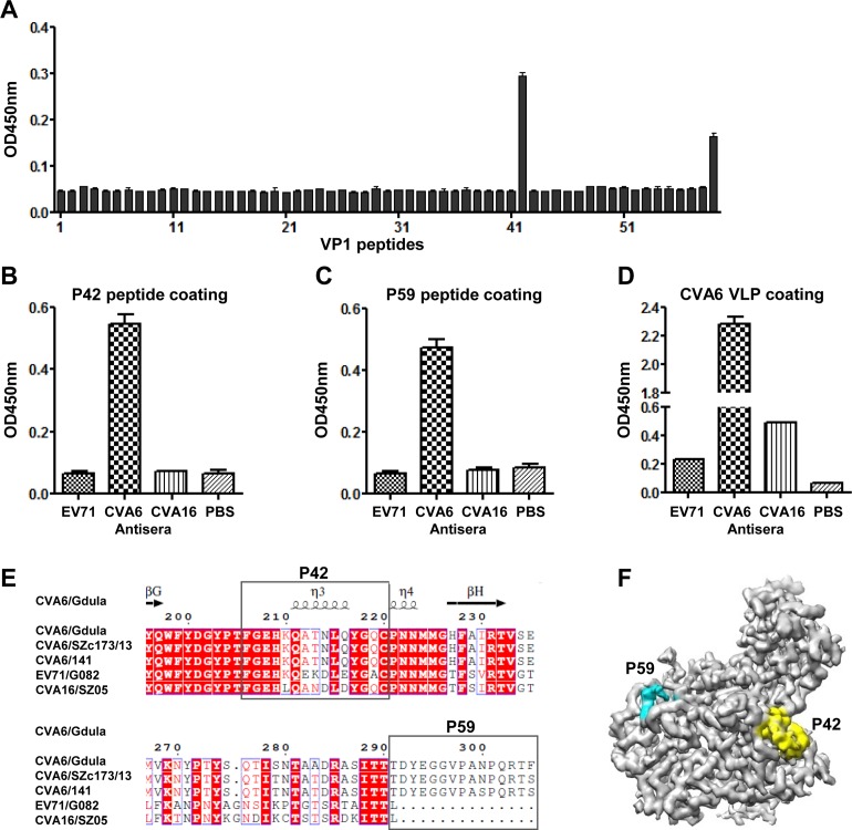
Coxsackievirus A6 (CVA6) has recently emerged as one of the predominant causative agents of hand, foot, and mouth disease (HFMD). The structure of the CVA6 mature viral particle has not been solved thus far. Our previous work shows that recombinant virus-like particles (VLPs) of CVA6 represent a promising CVA6 vaccine candidate. Here, we report the first cryo-electron microscopy (cryo-EM) structure of the CVA6 VLP at 3.0-Å resolution. The CVA6 VLP exhibits the characteristic features of enteroviruses but presents an open channel at the 2-fold axis and an empty, collapsed VP1 pocket, which is broadly similar to the structures of the enterovirus 71 (EV71) VLP and coxsackievirus A16 (CVA16) 135S expanded particle, indicating that the CVA6 VLP is in an expanded conformation. Structural comparisons reveal that two common salt bridges within protomers are maintained in the CVA6 VLP and other viruses of the Enterovirus genus, implying that these salt bridges may play a critical role in enteroviral protomer assembly. However, there are apparent structural differences among the CVA6 VLP, EV71 VLP, and CVA16 135S particle in the surface-exposed loops and C termini of subunit proteins, which are often antigenic sites for enteroviruses. By immunological assays, we identified two CVA6-specific linear B-cell epitopes (designated P42 and P59) located at the GH loop and the C-terminal region of VP1, respectively, in agreement with the structure-based prediction of antigenic sites. Our findings elucidate the structural basis and important antigenic sites of the CVA6 VLP as a strong vaccine candidate and also provide insight into enteroviral protomer assembly.IMPORTANCE Coxsackievirus A6 (CVA6) is becoming one of the major pathogens causing hand, foot, and mouth disease (HFMD), leading to significant morbidity and mortality in children and adults. However, no vaccine is currently available to prevent CVA6 infection. Our previous work shows that recombinant virus-like particles (VLPs) of CVA6 are a promising CVA6 vaccine candidate. Here, we present a 3.0-Å structure of the CVA6 VLP determined by cryo-electron microscopy. The overall architecture of the CVA6 VLP is similar to those of the expanded structures of enterovirus 71 (EV71) and coxsackievirus A16 (CVA16), but careful structural comparisons reveal significant differences in the surface-exposed loops and C termini of each capsid protein of these particles. In addition, we identified two CVA6-specific linear B-cell epitopes and mapped them to the GH loop and the C-terminal region of VP1, respectively. Collectively, our findings provide a structural basis and important antigenic information for CVA6 VLP vaccine development.

Keywords: coxsackievirus A6; cryo-EM; epitope; near-atomic-resolution structure; virus-like particle.


Figures

FIG 1
FIG 1
Cryo-EM image of CVA6 VLP and resolution evaluation of the cryo-EM map. (A) A representative cryo-EM image of the CVA6 VLP. Bar, 50 nm. (B) Power spectrum of the micrograph shown in panel A. The white dashed curve indicates that the image contains a signal at 3.2-Å resolution. (C) Resolution assessment of the cryo-EM reconstruction by Fourier shell correlation (FSC) at a criterion of 0.143. (D) Local resolution evaluation of the cryo-EM map by ResMap. The resolution color bar (in angstroms) is also labeled. (E) Central section of the cryo-EM map visualized along the 5-fold symmetry axis.
FIG 2
FIG 2
Overall structure of the CVA6 VLP. (A) Cryo-EM density map of the CVA6 VLP viewed along the icosahedral 2-fold symmetry axis. The map is colored radially. The color bar labels the corresponding radius from the center of the sphere (in angstroms). The mesa, propeller, and canyon are indicated by black arrows. (B) Atomic model of the CVA6 VLP viewed along the 2-fold axis. VP0, VP1, and VP3 are colored in green, blue, and red, respectively. The same color scheme is used throughout, unless otherwise indicated. (C) Good fit between the model and the cryo-EM map at an icosahedral asymmetric unit of the CVA6 VLP. The visualization location with respect to the complete CVA6 VLP map is illustrated in the upper left corner. (D) Good fit between the segmented density (mesh in gray) and the corresponding atomic model (sticks) in the VP1, VP0, and VP3 regions. The well-resolved density for almost all the side chains demonstrates the atomic resolution of the cryo-EM map.
FIG 3
FIG 3
Surface features of the CVA6 VLP compared to those of the EV71 VLP and CVA16 135S particle. (A) A map and model overlay viewed along the 2-fold symmetry axis (marked as a black ellipse) for the CVA6 VLP. The position of the junction channel is indicated by a black triangle. The density map is shown in transparent gray. (B and C) Atomic models of the outer surfaces of the EV71 VLP (PDB 4YVS) (B) and the CVA16 135S particle (PDB 4JGY) (C) viewed in the same direction as in panel A. (D to F) Surface representation of the pentameric structures of the CVA6 VLP (D), EV71 VLP (E), and CVA16 135S particle (F) viewed along the 5-fold symmetry axis. The surface is radially colored. The color bar labels the corresponding radius from the center of the particle (in angstroms). (G to I) Electrostatic surfaces of pentamers of the CVA6 VLP (G), EV71 VLP (H), and CVA16 135S particle (I). The electrostatic potential was calculated by utilizing PyMOL; the positively and negatively charged areas are shown in blue and red, respectively. The black dashed circle indicates the 5-fold positively charged patch.
FIG 4
FIG 4
Structural comparison of hydrophobic pockets among the CVA6 VLP, EV71 VLP, CVA16 135S particle, and EV71 mature virion. (A) The hydrophobic pocket in VP1 of the CVA6 VLP, with density in transparent gray and model in blue ribbon and stick. (B and C) Superposition of the VP1 pocket regions of the CVA6 VLP (blue), EV71 VLP (pink), CVA16 135S particle (cyan), and EV71 mature virion (PDB 3VBF) (orange) viewed from the front (B) and back (C). The pocket factor in the EV71 mature virion is shown as orange sticks. Shown also are residues M225 and F228 in the CVA6 VLP, whose side chains partially occupy the potential binding site for pocket factor.
FIG 5
FIG 5
Salt bridges within the protomers of the CVA6 VLP, EV71 VLP, and CVA16 135S particle. (A to C) Salt bridge locations within the protomers of the CVA6 VLP (A), EV71 VLP (B), and CVA16 135S particle (C). Salt bridge-forming residues within VP1, VP0/VP2 (VP0 for the CVA6 VLP and EV71 VLP and VP2 for the CVA16 135S particle), and VP3 are colored in blue, green, and red, respectively, while the other portions of the proteins are in gray. (D to F) Zoom-in view of the salt bridge regions enlarged from panels A, B, and C, respectively. The three common salt bridges observed in the CVA6 VLP, EV71 VLP, and CVA16 135S particle are indicated by oval dashed lines.
FIG 6
FIG 6
Structural comparison of the protomers and individual capsid proteins of the CVA6 VLP, EV71 VLP, and CVA16 135S particle. (A) Superposition of the protomers of the CVA6 VLP (different capsid proteins in different colors) and EV71 VLP (gray). The black oval, triangle, and pentagon represent the 2-fold, 3-fold, and 5-fold axes, respectively. (B) Superposition of the protomers of the CVA6 VLP and CVA16 135S particle (gray). VP1, VP0, and VP3 of CVA6 VLP are in blue, green, and red, respectively. (C to E) Superposition of individual capsid protein structures of the CVA6 VLP (blue), EV71 VLP (pink), and CVA16 135S particle (cyan). (C) Superposition of VP1. (D) Superposition of VP0/VP2 (VP0 for the CVA6 VLP and EV71 VLP and VP2 for the CVA16 135S particle). (E) Superposition of VP3. The major structural differences of subunit proteins among the CVA6 VLP, EV71 VLP, and CVA16 135S particle are indicated by black arrows. The dashed rectangle indicates the disordered region of the VP3 GH loop.
FIG 7
FIG 7
Identification and mapping of linear B-cell epitopes within the VP1 protein of CVA6. (A) Reactivity of the anti-CVA6 VLP mouse serum to VP1 peptides determined by peptide ELISA. A set of 59 synthetic peptides covering the entire VP1 region of CVA6 were used as the coating antigens. Mouse anti-CVA6 VLP serum was diluted 1:40 and used in ELISA. (B to D) Cross-reactivities of peptides P42 (B), P59 (C), and CVA6 VLP (D) with mouse polyclonal antibodies against the EV71 VLP or CVA16 VLP determined by ELISA. Anti-CVA6 VLP and anti-PBS mouse sera were used as positive and negative controls, respectively. The antisera were diluted 1:40 (B and C) and 1:100 (D) and used in ELISA. (E) Sequence alignment of VP1 from some representative strains of CVA6, EV71, and CVA16, showing the amino acid sequence variations in the corresponding P42 and P59 regions. The secondary-structure elements for the CVA6 VLP are shown at the top. (F) Locations of the P42 and P59 epitopes on the protomer of the CVA6 VLP. The P42 and P59 epitopes are colored in yellow and cyan, respectively. To better show the densities of P42 and P59, the map was low-pass filtered to 3.5-Å resolution. The electron densities of the nine C-terminal amino acids of P59 remain missing.

References

    1. Repass GL, Palmer WC, Stancampiano FF. 2014. Hand, foot, and mouth disease: identifying and managing an acute viral syndrome. Cleve Clin J Med 81:537–543. doi: 10.3949/ccjm.81a.13132. - DOI - PubMed
    1. Xing W, Liao Q, Viboud C, Zhang J, Sun J, Wu JT, Chang Z, Liu F, Fang VJ, Zheng Y, Cowling BJ, Varma JK, Farrar JJ, Leung GM, Yu H. 2014. Hand, foot, and mouth disease in China, 2008-12: an epidemiological study. Lancet Infect Dis 14:308–318. doi: 10.1016/S1473-3099(13)70342-6. - DOI - PMC - PubMed
    1. Wong SS, Yip CC, Lau SK, Yuen KY. 2010. Human enterovirus 71 and hand, foot and mouth disease. Epidemiol Infect 138:1071–1089. doi: 10.1017/S0950268809991555. - DOI - PubMed
    1. Bian L, Wang Y, Yao X, Mao Q, Xu M, Liang Z. 2015. Coxsackievirus A6: a new emerging pathogen causing hand, foot and mouth disease outbreaks worldwide. Expert Rev Anti Infect Ther 13:1061–1071. doi: 10.1586/14787210.2015.1058156. - DOI - PubMed
    1. Osterback R, Vuorinen T, Linna M, Susi P, Hyypia T, Waris M. 2009. Coxsackievirus A6 and hand, foot, and mouth disease, Finland. Emerg Infect Dis 15:1485–1488. doi: 10.3201/eid1509.090438. - DOI - PMC - PubMed

Publication types

MeSH terms

LinkOut - more resources