. 2018 Jan 30;92(4):e01628-17.
doi: 10.1128/JVI.01628-17. Print 2018 Feb 15.

Glycan Shield and Fusion Activation of a Deltacoronavirus Spike Glycoprotein Fine-Tuned for Enteric Infections

Affiliations

Glycan Shield and Fusion Activation of a Deltacoronavirus Spike Glycoprotein Fine-Tuned for Enteric Infections

Xiaoli Xiong et al. J Virol. .

Abstract

Coronaviruses recently emerged as major human pathogens causing outbreaks of severe acute respiratory syndrome and Middle East respiratory syndrome. They utilize the spike (S) glycoprotein anchored in the viral envelope to mediate host attachment and fusion of the viral and cellular membranes to initiate infection. The S protein is a major determinant of the zoonotic potential of coronaviruses and is also the main target of the host humoral immune response. We report here the 3.5-Å-resolution cryo-electron microscopy structure of the S glycoprotein trimer from the pathogenic porcine deltacoronavirus (PDCoV), which belongs to the recently identified Deltacoronavirus genus. Structural and glycoproteomics data indicate that the glycans of PDCoV S are topologically conserved compared with the human respiratory coronavirus NL63 S, resulting in similar surface areas being shielded from neutralizing antibodies and implying that both viruses are under comparable immune pressure in their respective hosts. The structure further reveals a shortened S2' activation loop, containing a reduced number of basic amino acids, which participates in rendering the spike largely protease resistant. This property distinguishes PDCoV S from recently characterized betacoronavirus S proteins and suggests that the S protein of enterotropic PDCoV has evolved to tolerate the protease-rich environment of the small intestine and to fine-tune its fusion activation to avoid premature triggering and reduction of infectivity.IMPORTANCE Coronaviruses use transmembrane S glycoprotein trimers to promote host attachment and fusion of the viral and cellular membranes. We determined a near-atomic-resolution cryo-electron microscopy structure of the S ectodomain trimer from the pathogenic PDCoV, which is responsible for diarrhea in piglets and has had devastating consequences for the swine industry worldwide. Structural and glycoproteomics data reveal that PDCoV S is decorated with 78 N-linked glycans obstructing the protein surface to limit accessibility to neutralizing antibodies in a way reminiscent of what has recently been described for a human respiratory coronavirus. PDCoV S is largely protease resistant, which distinguishes it from most other characterized coronavirus S glycoproteins and suggests that enteric coronaviruses have evolved to fine-tune fusion activation in the protease-rich environment of the small intestine of infected hosts.

Keywords: coronaviruses; cryo-EM; fusion proteins.


Figures

FIG 1
FIG 1
Cryo-EM structure of the PDCoV S protein. (A) A representative micrograph of vitreous ice-embedded PDCoV S protein at 3.4-μm defocus. Scale bar, 510 Å. (B) Selected 2D class averages of the PDCoV S protein. Scale bar, 85 Å. (C and D) Side (C) and top (D) views of the PDCoV S cryo-EM map filtered at 3.5-Å resolution and sharpened with a B-factor of −150 Å2. The density is colored for each protomer. (E and F) Ribbon representation of the PDCoV S trimer structure rendered with the same orientations as those in panels C and D. One protomer is colored according to the indicated structural domains, whereas the other two protomers are colored gray.
FIG 2
FIG 2
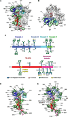
Glycosylation profile of the PDCoV S protein. (A and B) Two orthogonal views of the PDCoV S trimer rendered as ribbons. Glycan density extracted from the unsharpened reconstruction is colored green for one protomer and gray for the other two protomers. Labels indicate the position of N-linked glycosylated asparagine residues. (C) Schematic summary of all detected N-linked glycans. Each site shows the most extensive glycan structure detected, either by mass spectrometry or cryo-EM. A full overview of all detected N-linked glycans is provided in Table S1 in the supplemental material. Glycan moieties are represented as symbols according to the key, and the structural domains are individually colored and indicated in a linear representation of the PDCoV S sequence. (D and E) Ribbon representation of PDCoV (D) and HCoV-NL63 (E) S protomers with glycans visualized by cryo-EM shown as green spheres.
FIG 3
FIG 3
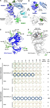
Structural features of the PDCoV S1 subunit and the galectin-like domain A. (A) Superposition of the PDCoV and HCoV-NL63 S1 subunits highlights the absence of domain 0 in PDCoV S. (B) View of the interface between PDCoV S A and B domains showing the Asn-184 glycan points away from domain B. (C) View of the interface between HCoV-NL63 S A and B domains showing the Asn-358 glycan contributes to masking the receptor-binding loops. (D) Ribbon representation of PDCoV domain A. (E) Ribbon representation of BCoV domain A oriented identically to panel D. Highly conserved residues involved in sialic acid recognition are shown in ball-and-stick representation. Glycans are rendered as spheres in panels A to C or sticks in panels D and E and colored by atom type (carbon, green; nitrogen, blue; oxygen, red). (F) The PDCoV S1 subunit C-terminally tagged with the Fc portion of human IgG (S1-Fc) was tested for its hemagglutination potential of an erythrocyte suspension of human or rat origin, either alone or premixed with protein A-coupled nanoparticles to increase the avidity of S1-Fc proteins for sialic acids. The sialic acid-binding S1 subunit of HCoV-OC43 (GenBank accession no. AAR01015.1) C-terminally fused to human Fc portion was used as a positive control. Mock indicates the condition where no S1 subunit was used (negative control). Wells positive for hemagglutination are encircled.
FIG 4
FIG 4
Structural comparison of α- and δ-coronavirus receptor-binding domains. (A to D) Ribbon rendering of the putative receptor-binding domain B of the δ-genus PDCoV S (A) and α-genus PRCV S (B), HCoV-NL63 S (C), and TGEV S (D). Loops that have been implicated in receptor binding for α-coronaviruses are indicated. Key aromatic residues that have been shown to take part in α-coronavirus receptor binding and putatively involved in δ-coronavirus receptor binding are highlighted. Disulfide bonds that stabilize receptor-binding loops are indicated, and glycans within the domain are shown as sticks (carbon, green; nitrogen, blue; oxygen, red).
FIG 5
FIG 5
Structural features of the PDCoV S2 subunit. (A) Ribbon representation of the PDCoV S trimer with the S2 subunit core of one protomer colored from blue to red (from N terminus to C terminus). (B) Zoomed-in view of the S2′ activation loop region. Two glycans, linked to Asn-669 and Asn-673, which are strictly conserved in HCoV-NL63 S, are shown as sticks (carbon, green; nitrogen, blue; oxygen, red). For comparison, the equivalent residues in the HCoV-NL63 S protein are indicated in gray. (C) The PDCoV S glycoprotein features an insertion of 14 amino acid residues in HR1 compared to the β-coronavirus MHV S protein, folding as an extended loop and a helical extension of two turns. The residues accounting for this HR1 insertion interact with the complementary insertion in HR2 in the postfusion conformation (see Fig. S2B).
FIG 6
FIG 6
PDCoV S glycoprotein is resistant to digestive enzymes. Purified SARS S (1 mg/ml) and PDCoV S (0.5 mg/ml) glycoproteins were incubated with 0.1 mg/ml trypsin or chymotrypsin for 2 h at 22°C. The digestion reactions were analyzed on a 12% SDS-PAGE gel. After incubation, the SARS S protein was extensively proteolyzed, whereas a large fraction of the PDCoV S protein remains intact.

References

    1. Zumla A, Chan JF, Azhar EI, Hui DS, Yuen KY. 2016. Coronaviruses–drug discovery and therapeutic options. Nat Rev Drug Discov 15:327–347. doi:10.1038/nrd.2015.37. - DOI - PMC - PubMed
    1. Vijay R, Perlman S. 2016. Middle East respiratory syndrome and severe acute respiratory syndrome. Curr Opin Virol 16:70–76. doi:10.1016/j.coviro.2016.01.011. - DOI - PMC - PubMed
    1. Schrauwen EJ, Fouchier RA. 2014. Host adaptation and transmission of influenza A viruses in mammals. Emerg Microbes Infect 3:e9. doi:10.1038/emi.2014.9. - DOI - PMC - PubMed
    1. Li F, Li W, Farzan M, Harrison SC. 2005. Structure of SARS coronavirus spike receptor-binding domain complexed with receptor. Science 309:1864–1868. doi:10.1126/science.1116480. - DOI - PubMed
    1. Lu G, Hu Y, Wang Q, Qi J, Gao F, Li Y, Zhang Y, Zhang W, Yuan Y, Bao J, Zhang B, Shi Y, Yan J, Gao GF. 2013. Molecular basis of binding between novel human coronavirus MERS-CoV and its receptor CD26. Nature 500:227–231. doi:10.1038/nature12328. - DOI - PMC - PubMed

Publication types

Substances