Clinical Trial
. 2017 Nov 2;2(21):e96381.
doi: 10.1172/jci.insight.96381.

Human vaccination against RH5 induces neutralizing antimalarial antibodies that inhibit RH5 invasion complex interactions

Affiliations
Clinical Trial

Human vaccination against RH5 induces neutralizing antimalarial antibodies that inhibit RH5 invasion complex interactions

Ruth O Payne et al. JCI Insight. .

Abstract

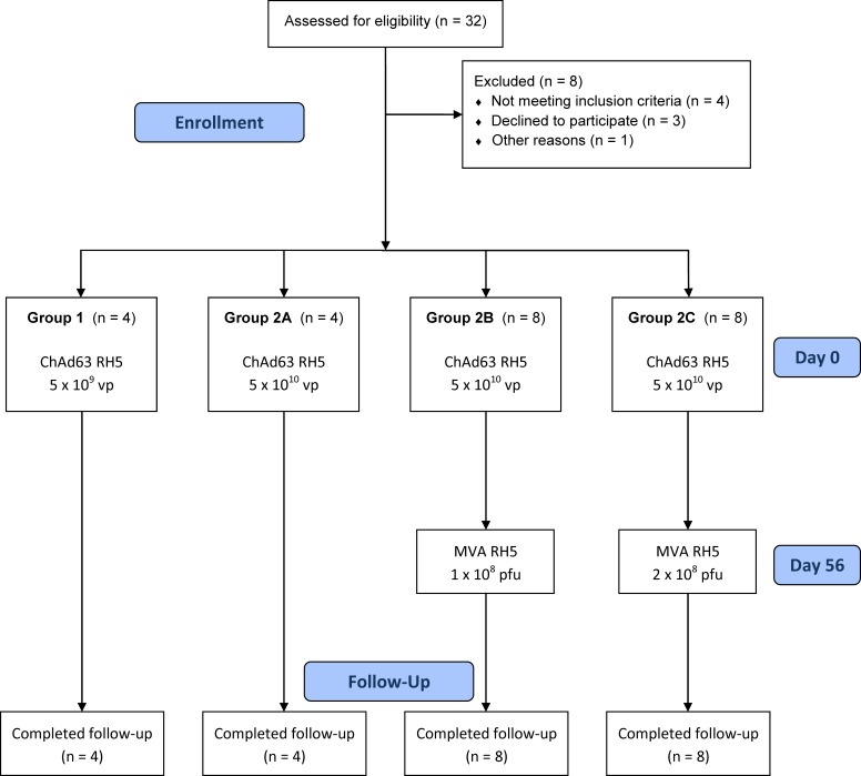
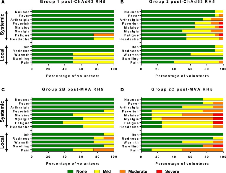
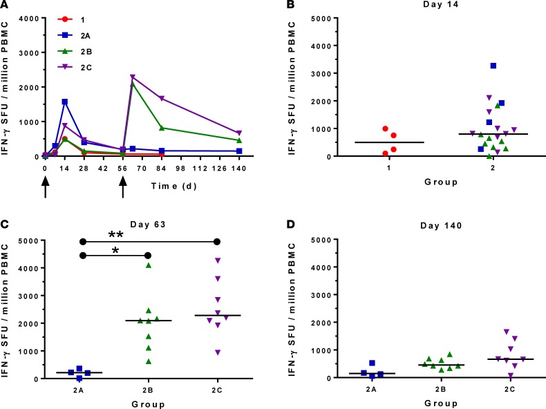
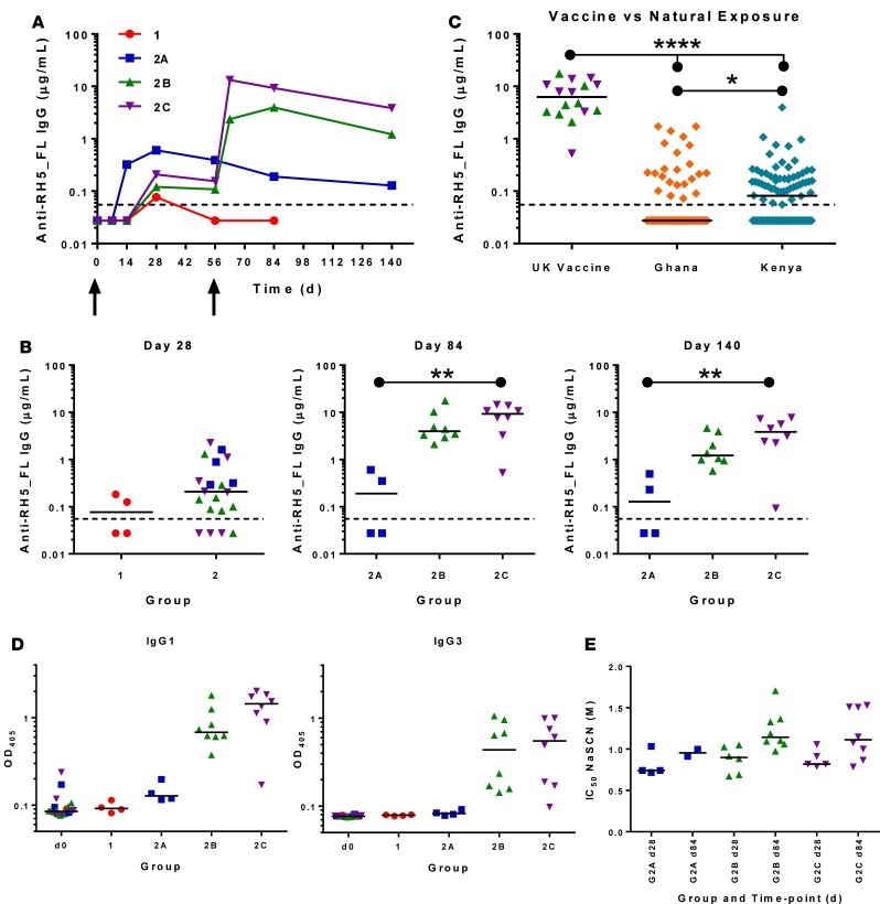
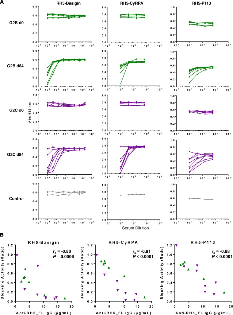
The development of a highly effective vaccine remains a key strategic goal to aid the control and eventual eradication of Plasmodium falciparum malaria. In recent years, the reticulocyte-binding protein homolog 5 (RH5) has emerged as the most promising blood-stage P. falciparum candidate antigen to date, capable of conferring protection against stringent challenge in Aotus monkeys. We report on the first clinical trial to our knowledge to assess the RH5 antigen - a dose-escalation phase Ia study in 24 healthy, malaria-naive adult volunteers. We utilized established viral vectors, the replication-deficient chimpanzee adenovirus serotype 63 (ChAd63), and the attenuated orthopoxvirus modified vaccinia virus Ankara (MVA), encoding RH5 from the 3D7 clone of P. falciparum. Vaccines were administered i.m. in a heterologous prime-boost regimen using an 8-week interval and were well tolerated. Vaccine-induced anti-RH5 serum antibodies exhibited cross-strain functional growth inhibition activity (GIA) in vitro, targeted linear and conformational epitopes within RH5, and inhibited key interactions within the RH5 invasion complex. This is the first time to our knowledge that substantial RH5-specific responses have been induced by immunization in humans, with levels greatly exceeding the serum antibody responses observed in African adults following years of natural malaria exposure. These data support the progression of RH5-based vaccines to human efficacy testing.


Conflict of interest statement

Conflict of interest: S.J. Draper is a named inventor on patent applications relating to RH5 and/or other malaria vaccines and immunization regimens; is a cofounder of, shareholder in, and consultant for SpyBiotech; and declares research funding support from Pfizer and GSK BioPharm. A.D. Douglas, G.J. Wright, and A.V.S. Hill are named inventors on patent applications relating to RH5 and/or other malaria vaccines and immunization regimens. L. Siani and S. Di Marco are employees of ReiThera (formerly Okairos), which is currently developing vectored vaccines for a number of diseases. J. Vekemans was an employee of GSK, which has acquired the ChAd63 vector from Okairos. R. Ashfield is a director of Ducentis and holds shares in the company, which is developing a therapy for autoimmune disease. A.M. Minassian has an immediate family member who is an inventor on patents relating to RH5 and/or other malaria vaccines and immunization regimens and who is a cofounder of, shareholder in, and consultant for SpyBiotech. S. Biswas is a cofounder and CEO of, and shareholder in, SpyBiotech and is a contributor in a patent application relating to multimerisation technology. J. Jin is a cofounder of and shareholder in SpyBiotech.

Figures

Figure 1
Figure 1. VAC057 flow chart of study design and volunteer recruitment.
Enrolment into the VAC057 study began in August 2014, and all follow-up visits were completed by October 28, 2015. All immunizations were administered i.m. into the deltoid of the nondominant arm preferentially.
Figure 2
Figure 2. Solicited AEs following vaccination with ChAd63 and MVA RH5.
The solicited local and systemic adverse events (AEs) recorded for 7 days following ChAd63 RH5 and MVA RH5 are shown at the maximum severity reported by all volunteers. (A) Four volunteers received 5 × 10⁹ viral particles (vp) ChAd63 RH5 (Group 1), and (B) 20 received 5 × 10¹⁰ vp (Group 2). (C) Eight of the Group 2 volunteers went on to receive MVA RH5 1 × 10⁸ plaque-forming units (pfu) (Group 2B), and (D) 8 received 2 × 10⁸ pfu (Group 2C).
Figure 3
Figure 3. Ex vivo IFN-γ T cell response to vaccination.
(A) Median ex vivo IFN-γ enzyme-linked immunospot (ELISPOT) responses in peripheral blood mononuclear cells (PBMC) to the RH5 insert (summed response across all the individual peptide pools) shown for all groups. Individual responses are shown in Supplemental Figure 1. Median and individual responses are shown at (B) d14 (n = 4 vs. 20); (C) d63 (G2A, n = 4; G2B, n = 8; G2C, n = 8) assessed by Kruskal-Wallis test with Dunn’s multiple comparison test; and (D) d140 (G2A n = 4, G2B n = 8, G2C n = 8). Symbols are coded according to group. *P < 0.05, **P < 0.01.
Figure 4
Figure 4. Serum antibody response to vaccination.
Group 1 (n = 4), Group 2A (n = 4), Group 2B (n = 8), Group 2C (n = 8). (A) Median anti–RH5_FL serum total IgG responses shown for all groups over time. Individual responses are shown in Supplemental Figure 3. Median and individual responses are shown at (B) d28, d84, and d140. The horizontal dotted line indicates the limit of detection of the assay. Statistical analysis using Kruskal-Wallis test with Dunn’s multiple comparison test. (C) Vaccine-induced responses shown for Groups 2B and 2C combined (n = 16) vs. responses following natural exposure in Ghanaian adults (n = 79) and Kenyan adults (n = 96); analysis by Kruskal-Wallis test with Dunn’s multiple comparison test. (D) Isotype profiles of serum antibody responses against RH5_FL were assessed by ELISA. Responses are shown at baseline (d0) and for all groups at d84. Individual and median responses are shown for IgG1 and IgG3; results for IgG2, IgG4, IgA, and IgM are shown in Supplemental Figure 5. (E) Avidity of serum IgG responses at d28 and d84 was assessed by sodium thiocyanate (NaSCN) displacement RH5_FL ELISA and is reported as the molar (M) concentration of NaSCN required to reduce the starting OD in the ELISA by 50% (IC50). Only samples with a positive response by anti–RH5_FL total IgG ELISA could be assayed for avidity. Symbols are coded according to group. *P < 0.05, **P < 0.01, ****P < 0.0001.
Figure 5
Figure 5. B cell response to vaccination.
(A) RH5-specific antibody-secreting cell (ASC) responses were assessed by ex vivo enzyme-linked immunospot (ELISPOT) using RH5_FL protein and fresh peripheral blood mononuclear cells (PBMC) from the d63 time point. Individual and median responses are shown for each group and reported as RH5-specific ASC per million PBMC used in the assay (n = 8 for Group 2B and n = 7 for Group 2C because 1 sample was not tested in this group). (B) Correlation of the ASC response vs. the concentrations of serum anti–RH5_FL IgG measured at d84. Spearman’s rank correlation coefficient (rs) and P value are shown by Spearman’s rank correlation. (C) RH5-specific memory B cell (mBC) responses were assessed by ELISPOT assay using RH5_FL protein (n = 8 for Groups 2B and 2C). Frozen PBMC were thawed and underwent a 6-day polyclonal restimulation, during which ASC are derived from mBC, before testing in the assay. Individual and median responses are shown from the d84 and 140 time points and are reported as mBC-derived RH5-specific ASC per million cultured PBMC or as (D) % of total number IgG-secreting ASC (n = 7 for Group 2C at the d140 time point in D, otherwise n = 8). Groups 2B and 2C are coded by color and symbol.
Figure 6
Figure 6. Functional GIA induced by ChAd63-MVA RH5 vaccination.
(A) In vitro growth inhibition activity (GIA) of purified IgG was assessed at 10 mg/ml against 3D7 clone P. falciparum parasites. Individual data and medians are shown for each group at d84 (G1, n = 4; G2A, n = 4; G2B, n = 8; G2C, n = 8); pooled sera were used for each group (n = 4) at baseline (d0). (B) Dilution series of purified IgG from Group 2B and 2C d84 samples. (C) Relationship between GIA data from the dilution series shown in B and concentration of anti–RH5_FL purified IgG used in the assay as measured by ELISA. The EC50 (concentration of anti-RH5_FL polyclonal IgG that gives 50% GIA, dashed line) was 8.2 μg/ml (95% CI, 7.2–9.5 μg/ml); nonlinear regression curve is shown (solid line, r2 = 0.90, n = 74). Two volunteers (1 in Group 2B and 1 in 2C) showed a reproducibly higher EC50 of 3.3 μg/ml (95% CI, 2.8–3.9 μg/ml); nonlinear regression curve is shown (dotted line, r2 = 0.99, n = 10). (D) Purified IgG from Group 2B and 2C d84 samples, plus 1 pooled d0 preimmunization sample, were tested at 10 mg/ml against a panel of 8 other laboratory-adapted parasite lines and short-term culture-adapted parasite isolates. GIA for each parasite and test sample is plotted against corresponding GIA against 3D7 clone parasites on the x axis.
Figure 7
Figure 7. Vaccine-induced anti-RH5 antibodies recognize linear epitopes and RH5Nt.
(A) D0 and d84 sera for volunteers in Groups 2B and 2C (n = 16) were diluted 1:100 and tested against linear overlapping peptides spanning the RH5 vaccine insert. Median, interquartile range (IQR), and range are shown for each peptide. (B) Plot of disorder within the RH5 vaccine construct predicted by PONDR. Blue arrows indicate the regions removed in the RH5ΔNL protein (E26-Y139 and N248-M296). (C) D0 and d84 sera for volunteers in Groups 2B (green triangles) and 2C (purple triangles) (n = 16) were diluted 1:100 and tested against 19-mer peptides that represent the minimal P113 binding region within RH5Nt (K33-K51). Peptides with N- and C-terminal biotinylation were tested to allow for binding to streptavidin-coated plated in both orientations. Individual and median results are shown. (D) D0 and d84 sera for volunteers in Groups 2B and 2C were diluted 1:100 and tested against RH5Nt protein. Individual and median results are shown (n = 16). (E) Correlation of d84 serum IgG responses in Groups 2B and 2C (n = 16) against RH5_FL and RH5Nt. Spearman’s rank correlation coefficient (rs) and P value are shown by Spearman’s rank correlation.
Figure 8
Figure 8. Vaccine-induced antibodies recognize conformational epitopes and inhibit interactions within the RH5 invasion complex.
(A) D0 and d84 sera for volunteers in Groups 2B and 2C (n = 16) were tested by ELISA against nondenatured RH5_FL protein (–) and the same protein following heat denaturation (+). Individual and median responses are shown. The 4BA7 and 2AC7 mAbs were included as controls that bind a linear vs. conformational epitope, respectively. **P < 0.01 according to Wilcoxon matched-pairs signed rank test. (B) D84 serum ELISA responses to RH5_FL and RH5ΔNL for volunteers in Groups 2B and 2C (n = 16) were analyzed for concordance by linear regression (solid line). r2 = 0.69; slope = 0.91 (95% CI, 0.56–1.27); Y intercept when X = 0.0 is 0.4 (95% CI, –2.7–3.6); X intercept when Y = 0.0 is –0.5 (95% CI, –6.0–2.3). Line of identity (X=Y) is also shown (dashed line).
Figure 9
Figure 9. Vaccine-induced antibodies inhibit interactions within the RH5 invasion complex.
(A) D0 and d84 Group 2B sera (n = 7) and Group 2C sera (n = 8) were tested for their ability to inhibit the interaction between proteins from the RH5 invasion complex by AVEXIS. Dilution of each test serum sample is shown starting at 1:10. Results with various assay controls also shown (no serum for RH5-Basigin, and anti-AMA1 for RH5-CyRPA and RH5-P113). Each point represents the mean of duplicate or triplicate wells. (B) Correlation of blocking activity for each interaction using d84 sera from Groups 2B and 2C (n = 15) against anti–RH5_FL serum IgG responses measured by ELISA. Blocking activity was calculated for each individual sample from the data in panel A as the ratio of the Abs 485 nm at 1:10 serum dilution divided by the Abs 485 nm at the highest serum dilution tested. Spearman’s rank correlation coefficient (rs) and P value(Spearman’s rank correlation) are shown.

References

    1. WHO. World Malaria Report 2015. Geneva, Switzerland: World Health Organization; 2015. http://www.who.int/malaria/publications/world-malaria-report-2015/report... Accessed October 10, 2017.
    1. Moorthy VS, Newman RD, Okwo-Bele JM. Malaria vaccine technology roadmap. Lancet. 2013;382(9906):1700–1701. - PubMed
    1. Halbroth BR, Draper SJ. Recent developments in malaria vaccinology. Adv Parasitol. 2015;88:1–49. - PubMed
    1. Efficacy and safety of RTS,S/AS01 malaria vaccine with or without a booster dose in infants and children in Africa: final results of a phase 3, individually randomised, controlled trial. Lancet. 2015;386(9988):31–45. doi: 10.1016/S0140-6736(15)60721-8. RTS,S Clinical Trials Partnership. - DOI - PMC - PubMed
    1. Sissoko MS, et al. Safety and efficacy of PfSPZ Vaccine against Plasmodium falciparum via direct venous inoculation in healthy malaria-exposed adults in Mali: a randomised, double-blind phase 1 trial. Lancet Infect Dis. 2017;17(5):498–509. doi: 10.1016/S1473-3099(17)30104-4. - DOI - PMC - PubMed

Publication types