. 2017 Nov 1;7(1):14803.
doi: 10.1038/s41598-017-14790-2.

Retinoic acid signaling maintains epithelial and mesenchymal progenitors in the developing mouse ureter

Affiliations

Retinoic acid signaling maintains epithelial and mesenchymal progenitors in the developing mouse ureter

Tobias Bohnenpoll et al. Sci Rep. .

Abstract

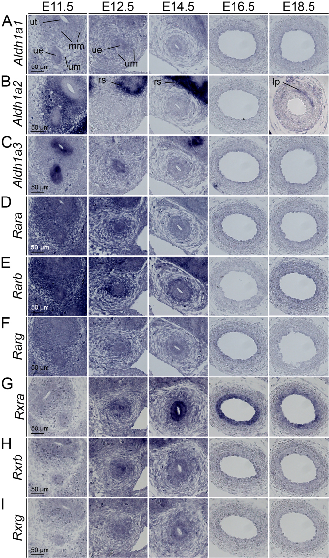
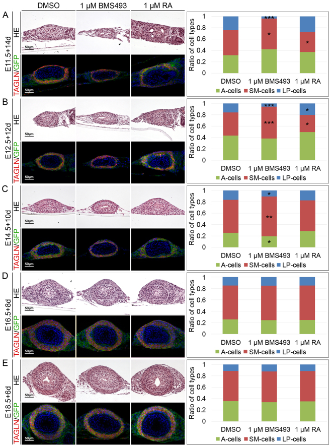
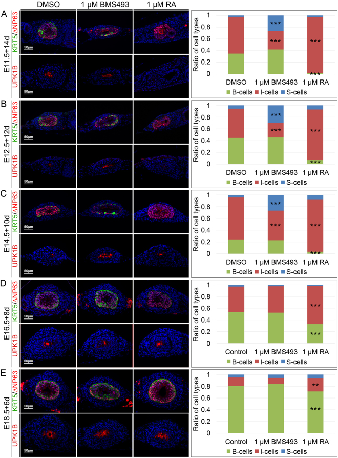
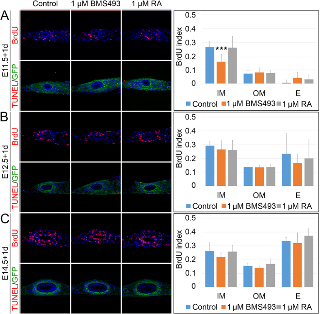
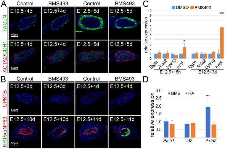
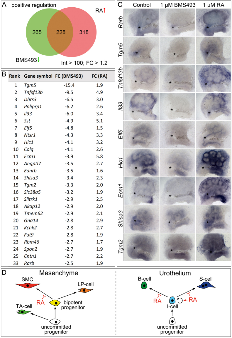
The differentiated cell types of the mature ureter arise from the distal ureteric bud epithelium and its surrounding mesenchyme. Uncommitted epithelial cells first become intermediate cells from which both basal and superficial cells develop. Mesenchymal progenitors give rise to separated layers of adventitial fibrocytes, smooth muscle cells and lamina propria fibrocytes. How progenitor expansion and differentiation are balanced is poorly understood. Here, we addressed the role of retinoic acid (RA) signaling in these programs. Using expression analysis of components and target genes, we show that pathway activity is restricted to the mesenchymal and epithelial progenitor pools. Inhibition of RA signaling in ureter explant cultures resulted in tissue hypoplasia with a relative expansion of smooth muscle cells at the expense of lamina propria fibroblasts in the mesenchyme, and of superficial cells at the expense of intermediate cells in the ureteric epithelium. Administration of RA led to a slight reduction of smooth muscle cells, and almost completely prevented differentiation of intermediate cells into basal and superficial cells. We identified cellular programs and transcriptional targets of RA signaling that may account for this activity. We conclude that RA signaling is required and sufficient to maintain mesenchymal and epithelial progenitors in early ureter development.


Conflict of interest statement

The authors declare that they have no competing interests.

Figures

Figure 1
Figure 1
RA signaling is active in early ureter development. In situ hybridization analysis on transverse sections of the proximal ureter of wild-type embryos for expression of genes encoding RA synthesizing genes (AC), RA receptors (DF) and retinoid X receptors (GI). Stages and probes are as indicated. For explanations see main text. lp, lamina propria; mm, metanephrogenic mesenchyme; rs, renal stroma; ue, ureteric epithelium; um, ureteric mesenchyme; ut, ureter tip.
Figure 2
Figure 2
RA signaling controls ureteric mesenchymal differentiation. Hematoxylin and eosin (upper panel) and co-immunofluorescence analysis of GFP and TAGLN (lower panel) on transverse proximal sections of E11.5 + 14d (A), E12.5 + 12d (B), E14.5 + 10d (C), E16.5 + 8d (D) and E18.5 + 6d (E) Tbx18 cre/+; R26 mTmG/+ ureter explants that were treated with DMSO, 1 µM BMS493 or 1 µM RA. The bar graph displays ratios of differentiated cell types that were quantified based on the immunofluorescence analysis. RA signaling acts from E11.5 to E14.5 to maintain lamina propria cells at the expense of SMCs. For numbers and statistics see Supplementary Table S1. *p ≤ 0.01; **p ≤ 0.001; ***p ≤ 0.0001.
Figure 3
Figure 3
RA signaling controls urothelial differentiation. Co-immunofluorescence analysis of KRT5 and ∆NP63 (upper panel) and UPK1B (lower panel) on transverse proximal sections of E11.5 + 14d (A), E12.5 + 12d (B), E14.5 + 10d (C), E16.5 + 8d (D) and E18.5 + 6d (E) wildtype ureter explants that were treated with DMSO, 1 µM BMS493 or 1 µM RA. The bar diagram displays ratios of differentiated cell types that were quantified based on the immunofluorescence analysis. RA signaling acts from E11.5 to E14.5 to maintain I-cells at the expense of S-cells. Throughout all stages analyzed ectopic RA prevents the differentiation of B-cells. For numbers and statistics see Supplementary Table S2. **p ≤ 0.001; ***p ≤ 0.0001.
Figure 4
Figure 4
RA signaling affects proliferation in a minor way. Determination of cellular proliferation by BrdU incorporation assay (upper row) and apoptosis by TUNEL/GFP assay (lower row) on transverse sections of the proximal ureter of E11.5 (A), E12.5 (B) and E14.5 (C) Tbx18 cre/+ ; R26 mTmG/+ explants that were cultured in the presence of DMSO, 1 µM BMS493 or 1 µM RA for 1 day. RA signaling is required to maintain proliferation of inner mesenchymal cells at E11.5. The bar diagrams display the BrdU incorporation indices in arbitrarily defined compartments of the ureter under given conditions. For numbers and statistics see Supplementary Table S3. ***p ≤ 0.0001. E, epithelium; IM, inner mesenchyme; OM, outer mesenchyme.
Figure 5
Figure 5
RA signaling prevents precocious differentiation in the ureter. (A) Immunofluorescence analysis for TAGLN (upper row) and co-immunofluorescence analysis of ACTA/CDH1 (lower row) on E12.5 ureter explants that were cultured for 3 and 4 days in presence of DMSO or 1 µM BMS493 shows that SMCs markers are slightly enhanced in their expression when RA signaling is abrogated. (B) Immunofluorescence analysis for UPK1B (upper row) and co-immunofluorescence analysis of KRT5/∆NP63 (lower row) on E12.5 ureter explants that were cultured for 3 and 4 days (UPK1B) or 10 and 11 days (KRT5/∆NP63) in the presence of DMSO or 1 µM BMS493. Markers for S-cells (UPK1B) and B-cells (KRT5) are prematurely expressed when RA signaling is inhibited. (C) RT-PCR analysis of expression of the SMC marker Tagln and Acta2, the S-cell marker Upk1b and the B-cell marker Krt5 in ureters treated for 18 h and 3 days with DMSO or the RA signaling inhibitor BMS493. Note the strong increase of expression of Krt5 at both stages. (D) RT-PCR analysis of expression of the SHH target gene Ptch1, of the WNT target gene Axin2 and the BMP target gene Id2 in ureters treated for 18 h with DMSO, 1 µM BMS493 or 1 µM RA. Note increase of expression of Axin2 almost reaching significance as indicated by (*). For numbers and statistics (for C and D) see Supplementary Table S4 and 5. *p ≤ 0.01; **p ≤ 0.001.
Figure 6
Figure 6
Identification of genes positively regulated by RA signaling in microarray experiments. Summary of the results from the microarray analysis of E12.5 ureters explanted and treated with DMSO or 1 µM BMS493 or 1 µM RA for 18 h filtered with an intensity (Int) threshold of 100 and a fold change (FC) cut-off of 1.2. (A) The Venn diagram displays transcripts which were positively regulated by RA signaling, i.e. down-regulated upon BMS493 treatment (493, green) or up-regulated upon RA treatment (546, red). The intersection shows the common group of positively regulated transcripts (228). (B) Top 25 of the common positively regulated genes were ranked according to their FC down-regulation upon BMS493 treatment. (C) In situ hybridization analysis of expression of selected genes which were positively regulated by RA signaling on E11.5 kidney explant cultures, treated with 1 µM BMS493 or 1 µM RA for 1 day. (D) Schematic illustration of RA signaling function in the maintenance of ureteric progenitors. In the ureteric mesenchyme (left) uncommitted progenitors diversify into adventitial fibrocytes (AF-cells) and bipotent progenitors of SMCs and lamina propria fibrocytes (LP-cells). RA specifically acts in the bipotent progenitors to prevent precocious differentiation towards the SMC lineage. In the ureteric epithelium (right) uncommitted progenitors give rise to I-cells which are maintained by RA signaling by preventing precocious differentiation towards the B- and S-cell lineage.

References

    1. Velardo, J. T. In The ureter (ed. H. Bergman. (Springer-Verlag, 1981).
    1. Bohnenpoll T, et al. Diversification of Cell Lineages in Ureter Development. J Am Soc Nephrol. 2017;28:1792–1801. doi: 10.1681/ASN.2016080849. - DOI - PMC - PubMed
    1. Yu J, Carroll TJ, McMahon AP. Sonic hedgehog regulates proliferation and differentiation of mesenchymal cells in the mouse metanephric kidney. Development. 2002;129:5301–5312. - PubMed
    1. Miyazaki Y, Oshima K, Fogo A, Ichikawa I. Evidence that bone morphogenetic protein 4 has multiple biological functions during kidney and urinary tract development. Kidney Int. 2003;63:835–844. doi: 10.1046/j.1523-1755.2003.00834.x. - DOI - PubMed
    1. Mamo TM, et al. BMP4 uses several different effector pathways to regulate proliferation and differentiation in the epithelial and mesenchymal tissue compartments of the developing mouse ureter. Hum Mol Genet. 2017;26:3553–3563. doi: 10.1093/hmg/ddx242. - DOI - PubMed

Publication types

MeSH terms

LinkOut - more resources