. 2017 Nov 1;7(1):14804.
doi: 10.1038/s41598-017-14208-z.

HtrA1 activation is driven by an allosteric mechanism of inter-monomer communication

Affiliations

HtrA1 activation is driven by an allosteric mechanism of inter-monomer communication

Alvaro Cortes Cabrera et al. Sci Rep. .

Abstract

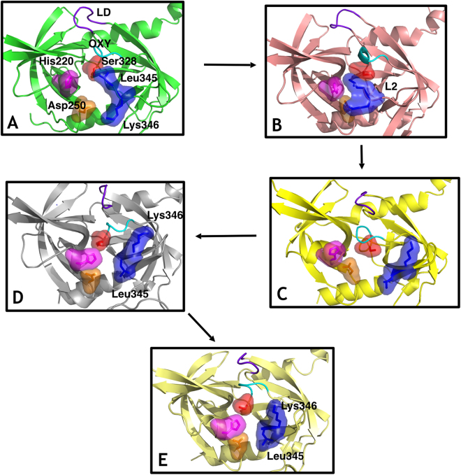
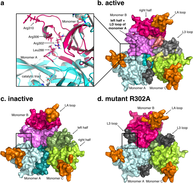
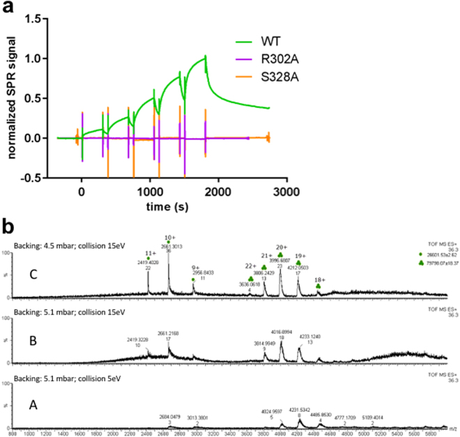
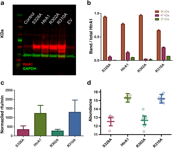
The human protease family HtrA is responsible for preventing protein misfolding and mislocalization, and a key player in several cellular processes. Among these, HtrA1 is implicated in several cancers, cerebrovascular disease and age-related macular degeneration. Currently, HtrA1 activation is not fully characterized and relevant for drug-targeting this protease. Our work provides a mechanistic step-by-step description of HtrA1 activation and regulation. We report that the HtrA1 trimer is regulated by an allosteric mechanism by which monomers relay the activation signal to each other, in a PDZ-domain independent fashion. Notably, we show that inhibitor binding is precluded if HtrA1 monomers cannot communicate with each other. Our study establishes how HtrA1 trimerization plays a fundamental role in proteolytic activity. Moreover, it offers a structural explanation for HtrA1-defective pathologies as well as mechanistic insights into the degradation of complex extracellular fibrils such as tubulin, amyloid beta and tau that belong to the repertoire of HtrA1.


Conflict of interest statement

All authors are F. Hoffman-La Roche employees or collaborators.

Figures

Figure 1
Figure 1
Sequence of states in the activation process from computational simulation. The residues Ser328 (surface, red), His220 (surface, magenta), Asp250 (surface, orange), L2 loop residues Leu345 and Lys346 (surface, blue), the LD loop (purple) and the oxyanion hole forming loop (cyan) are highlighted. (A) Fully inactive state that closely resembles the X-ray inactive structure (PDB 3NUM). The misaligned configuration of the catalytic triad (Ser328, His220 and Asp250) is incompatible with catalytic activity. In the binding pocket, the oxyanion hole-forming loop is disorganized and not functional while residue Leu345 occupies the cavity as a gate occluding the entrance. (B) The L2 loop adopts an intermediate conformation, where the side chain of the residues Leu345 and Lys346 are not opposed but parallel, while the rest of the elements involved in the catalytic mechanism remains similar to the inactive state A. (C) Leu345 and Lys346 have evolved to an active position from the disorganized intermediate B, inverting the orientation of their side chains originally displayed in their active conformations. This movement unblocks the S1 site of the protease while the remaining elements (catalytic triad, oxyanion hole forming loop and L3 loop) are in an inactive-like state as in state A and B. (D) The catalytic triad and all the remaining elements are now aligned in an active configuration. Only the oxyanion hole-forming loop remains disordered and not functional. (E) The structure is completely active and resembles the crystallographic active conformation (PDB 3NZI). Loop L3 stabilizes to adopt a well-defined secondary structure.
Figure 2
Figure 2
The dynamic communities of the HtrA1 trimeric protein are shown in different colors. (a) Intra-monomer community in the active trimer. L3 loop residues (Arg302-Arg310) of monomer B and LD residues (284–290) from monomer A belong to the same dynamic community. (b) Active trimer form. (c) Inactive trimer form. (d) Mutant R302A trimer form. Black regions in (b) and (d) indicate L3 loop independent communities.
Figure 3
Figure 3
(a) Surface plasmon resonance of HtrA1 variants binding to DPMFKL-boro-V: Wild-type HtrA1 catalytic domain (green), mutant R302A (purple) and S328A (dead domain). No binding is observed for R302A or S328A. (b) Native mass-spectrometry of HtrA1 mutant R302A. Peaks with three and one green dots correspond to trimer and monomer species respectively. At high-backing pressure (panel b.A) mostly trimer species is observed (ratios for WT r = ~6/2 = ~3, R302A r = ~8/3 = ~2.7, and S328A r = ~20/5 = ~4.4) whereas increasing collision energy and/or lowering backing pressure results in an increase in monomeric species (panels b.B and b.C) and decrease of the ratio r (Δr = 0.66 WT, Δr = 0.76 R302, Δr = 0.6). Also in this sample, higher collision energy or backing pressure increase m/z ratio indicating a slight loss of native folded trimeric state. (See Supp. Info. for native MS of wild-type and S328A).
Figure 4
Figure 4
(a) Western Blot (WB) of control sample (no virus), and vectors containing: HtrA1 mutant S328A (catalytic inactive control), HtrA1 wild-type, HtrA1 R302A mutant, HtrA1 R310A single mutant and empty vector. The yellow arrow indicates the 47 kDa band corresponding to the HtrA1 self-cleavage product. This image was cropped from an original blot displayed in Supporting Information. (b) WB quantification normalized first to GAPDH (Glyceraldehyde-3-phosphate dehydrogenase) and then to the total HtrA1 amount. Bands at 37, 47 and 54 kDa correspond to GAPDH, HtrA1 self-cleavage product and HtrA1 monomer respectively. (c) HtrA1 in vitro enzymatic assay measured in supernatant from WB, and normalized by total HtrA1 amount (Supp. Figure S5). (d) Extracellular HtrA1 activity monitored by the quantification of the semi-tryptic peptide NEQFNWVSR, which is generated through cleavage of clusterin at EQL362-NEQ.

References

    1. Dobson CM. Protein folding and misfolding. Nature. 2003;426:884–890. doi: 10.1038/nature02261. - DOI - PubMed
    1. Clausen T, Kaiser M, Huber R, Ehrmann M. HTRA proteases: regulated proteolysis in protein quality control. Nat Rev Mol Cell Biol. 2011;12:152–162. doi: 10.1038/nrm3065. - DOI - PubMed
    1. Tennstaedt A, et al. Human high temperature requirement serine protease A1 (HTRA1) degrades tau protein aggregates. J Biol Chem. 2012;287:20931–20941. doi: 10.1074/jbc.M111.316232. - DOI - PMC - PubMed
    1. Chien J, He X, Shridhar V. Identification of tubulins as substrates of serine protease HtrA1 by mixture‐based oriented peptide library screening. Journal of cellular biochemistry. 2009;107:253–263. doi: 10.1002/jcb.22121. - DOI - PMC - PubMed
    1. Dewan A, et al. HTRA1 promoter polymorphism in wet age-related macular degeneration. Science. 2006;314:989–992. doi: 10.1126/science.1133807. - DOI - PubMed

Publication types

MeSH terms