. 2017 Oct 18:8:1341.
doi: 10.3389/fimmu.2017.01341. eCollection 2017.

RANTES/CCL5 Induces Collagen Degradation by Activating MMP-1 and MMP-13 Expression in Human Rheumatoid Arthritis Synovial Fibroblasts

Affiliations

RANTES/CCL5 Induces Collagen Degradation by Activating MMP-1 and MMP-13 Expression in Human Rheumatoid Arthritis Synovial Fibroblasts

Solomon A Agere et al. Front Immunol. .

Abstract

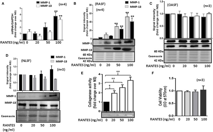
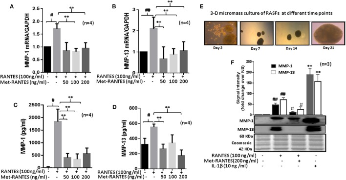
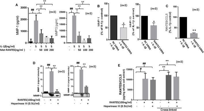
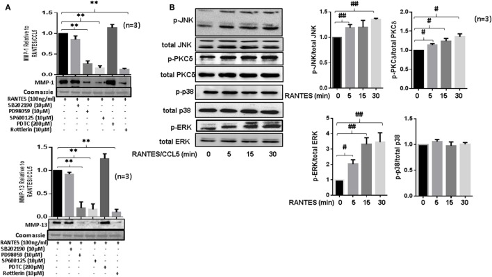
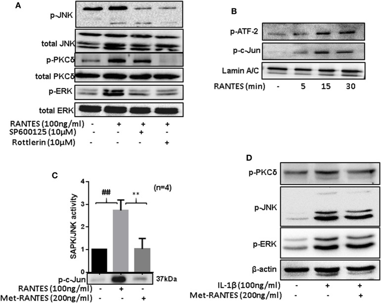
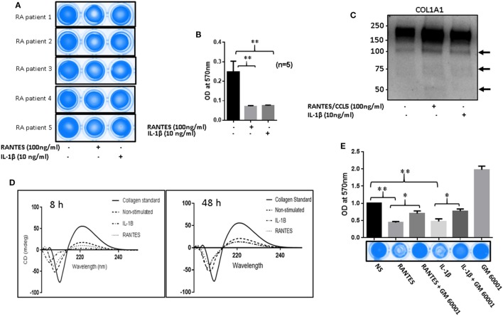
Regulated on activation, normal T expressed, and secreted (RANTES)/CC ligand 5 (CCL5) participates in rheumatoid arthritis (RA) pathogenesis by facilitating leukocyte infiltration, however, its other pathological functions are not fully defined in RA. In the present study, we evaluated the effect of RANTES/CCL5 on tissue degrading enzymes matrix metalloproteinase-1 (MMP-1) and MMP-13 expression and its contribution to the progressive joint damage by RA synovial fibroblasts (RASFs). Our results showed that RANTES/CCL5 dose dependently induced MMP-1 and MMP-13 expression in monolayers and three-dimensional (3D) micromass of human RASFs, which correlated with an increase in collagenase activity. This activation by RANTES/CCL5 was observed in RASF, but not in osteoarthritis SFs (OASFs). Evaluation of the signaling events showed that RANTES/CCL5 selectively activated PKCδ, JNK, and ERK proteins to induce MMP expression in human RASFs. Pretreatment with a functional antagonist (Met-RANTES) or heparinase III [an enzyme that selectively digests heparan sulfate proteoglycans (HSPGs)] completely abrogated RANTES/CCL5-induced MMP-1 and MMP-13 expression. Interestingly, the inhibition of RANTES/CCL5 using small-interfering RNA approach reduced the ability of interleukin-1β (IL-1β) to induce MMP-1 and MMP-13 expression, asserting its mediatory role in tissue remodeling. In the inhibitor study, only the selective inhibition of HSPGs or PKCδ, ERK, and JNK markedly inhibited RANTES/CCL5-induced MMP-1 and MMP-13 production. Circular dichroism spectroscopy results demonstrated the degradation of collagen triple-helical structure upon exposure to the conditioned media from RANTES/CCL5 stimulated RASFs, which was reverted by a broad-spectrum MMP inhibitor (GM6001). These findings suggest that RANTES/CCL5 not only upregulates MMP-1 and MMP-13 expression by partly utilizing HSPGs and/or PKCδ-JNK/ERK pathways but also mediates IL-1β-induced MMP-1 and MMP-13 expression.

Keywords: heparan sulfate proteoglycans; matrix metalloproteinases; normal T expressed; regulated on activation; rheumatoid arthritis; secreted/CC ligand 5; synovial fibroblasts.


Figures

Figure 1
Figure 1
Regulated on activation, normal T expressed, and secreted (RANTES)/CC ligand 5 (CCL5) induces matrix metalloproteinase (MMP)-1 and MMP-13 expression in rheumatoid arthritis synovial fibroblasts (RASFs). (A) Effect of RANTES/CCL5 on MMP-1 and MMP-13 mRNA and (B) protein expression was studied in RASFs. (C,D) Human OASFs and NLSFs were treated with different concentrations of recombinant RANTES/CCL5 (20, 50, and 100 ng/ml) and MMP-1 and MMP-13 expression was analyzed using Western immunoblotting, respectively. Gels described for (B–D) were stained with Commassie Blue stain to ensure equal loading of the supernatants. (E) Effect of RANTES/CCL5 on collagenase activity in RASFs. RASFs were treated with different concentration (20, 50, and 100 ng/ml) of RANTES/CCL5 for 24 h and conditioned media was concentrated using Amicon® Ultra Centrifugal filters (Millipore) and resolved on polyacrylamide gel loaded with collagen. Zymography image of digested regions representing MMPs activity is shown. Fold changes on developed zymograms was determined using densitometric analysis. (F) Effect of RANTES/CCL5 on the cell viability of RASFs was determined using MTT assay. RASFs were treated with RANTES/CCL5 (20, 50, and 100 ng/ml) for 24 h. Cell viability of cultured RASFs in the presence of RANTES/CCL5 was measured by optical densities at 570 nm. Values are represented as mean ± SE from three to four independent experiments using cells from different donors under similar conditions. *p < 0.05; **p < 0.01 for NS versus RANTES/CCL5 treatment.
Figure 2
Figure 2
Met-RANTES inhibits regulated on activation, normal T expressed, and secreted (RANTES)/CC ligand 5 (CCL5)-induced matrix metalloproteinase (MMP)-1 and MMP-13 expression in rheumatoid arthritis synovial fibroblasts (RASFs). (A,B) Effect of Met-RANTES on RANTES/CCL5-induced MMP-1 and MMP-13 mRNA and (C,D) at protein levels was determined using qRT-PCR and ELISA, respectively. RASFs were pretreated with Met-RANTES (50, 100, and 200 ng/ml) for 30 min followed by stimulation with RANTES/CCL5 (100 ng/ml) for 24 h. (E,F) Human RASFs were cultured in three-dimensional (3D) micromass for 21 days to form synovial tissue-like architect, followed by pretreatment with or without Met-RANTES for 30 min and then stimulation with RANTES (100 ng/ml) for 24 h. Interleukin (IL)-1β (10 ng/ml) 24 h stimulated RASF micromass were used as the positive control. Conditioned media was concentrated using Amicon® Ultra Centrifugal filters (Millipore) and MMP-1 and MMP-13 production was analyzed using Western immunoblotting. Densitometry was performed to determine the relative changes. Values are represented as mean ± SE from three to four independent experiments using cells from different donors. #p < 0.05 or ##p < 0.01 for NS vs. RANTES/CCL5 or IL-1β; **p < 0.01 for RANTES/CCL5 vs. Met-RANTES treatment. Representative figures of RASFs on 3D micromass culture (4× magnification; size 200–400 µm) are shown on days 2, 7, 14, and 21.
Figure 3
Figure 3
Regulated on activation, normal T expressed, and secreted (RANTES)/CC ligand 5 (CCL5) contributes to the proinflammatory cytokine-mediated matrix metalloproteinase (MMP)-1 and MMP-13 expression in rheumatoid arthritis (RA). (A) Met-RANTES inhibits interleukin (IL)-1β-induced MMP-1 and MMP-13 production in rheumatoid arthritis synovial fibroblasts (RASFs). (B) Effect of RANTES/CCL5 knockdown on IL-1β-induced MMP-1 and MMP-13 production in RASFs. RASFs were transfected with RANTES/CCL5-specific small-interfering RNA (siRNA) for 48 h and then stimulated with IL-1β (5 ng/ml) for 24 h. Levels of MMP-1 and MMP-13 in conditioned media was quantified using ELISA and represented as percent of IL-1β-treatment control. (C) Conditioned media from the above treated samples were also evaluated to determine percent knockdown of RANTES/CCL5 using siRNA approach and represented as percent of IL-1β treatment control. (D) Degradation of heparan sulfate proteoglycans (HSPG) by heparinase III reduces RANTES/CCL5-induced MMP-1 and MMP-13 expression in RASFs. RASFs were pretreated with heparinase III (0.5 U/ml) for 2 h followed by stimulation with RANTES/CCL5 (100 ng/ml) for 24 h. Conditioned media was concentrated and used to detect MMP-1 and MMP-13 expression using Western immunoblotting. Densitometry was performed to determine the relative changes. (E) RASFs pretreated with heparinase III (0.5 U/ml) for 2 h followed by stimulation with RANTES/CCL5 (100 ng/ml) for 1 h were used with or without cross-linking step for the detection of RANTES/CCL5 in the whole cell lysates (25 µl). Values are represented as mean ± SE from three independent experiments performed using RASFs from different donors under similar conditions. ##p < 0.01 for NS vs. or RANTES/CCL5 or IL-1β. *p < 0.05 or **p < 0.01 for RANTES/CCL5 or IL-1β vs. Met-RANTES or heparinase III treatment.
Figure 4
Figure 4
Regulated on activation, normal T expressed, and secreted (RANTES)/CC ligand 5 (CCL5) selectively activates PKCδ and JNK pathways to induce matrix metalloproteinase (MMP)-1 and MMP-13 production in rheumatoid arthritis synovial fibroblast (RASFs). (A) RASFs were pretreated with p-38 inhibitor (SB203980; 10 µM), ERK inhibitor (PD98059; 10 µM), JNK inhibitor (SP600125; 10 µM), NF-KB inhibitor (PDTC; 200 µM), or PKCδ inhibitor (Rottlerin; 10 µM) for 2 h followed by stimulation with RANTES/CCL5 for 24 h. Culture supernatant were concentrated to determine MMP-1 and MMP-13 expression using Western immunoblotting. Densitometry was performed to determine the relative changes. Gels were stained with Commassie Blue R250 stain to ensure equal loading. (B) Effect of RANTES/CCL5 (100 ng/ml) on the MAPK (p38, JNK and ERK) and PKCδ pathways was studied in human RASFs. RASFs were treated with RANTES (100 ng/ml) for different time (5, 15, and 30 min) and effect on phosphorylation of p38, JNK, ERK, and PKCδ was determined using Western immunoblotting. Total forms of p-38, JNK, ERK, and PKCδ were used as normalizing controls, respectively. Densitometry was performed to show the relative changes. Values are represented as mean ± SE from three or more independent experiments using cells from different donors under similar conditions. **p < 0.01 for RANTES/CCL5 vs. inhibitors; #p < 0.05 or ##p < 0.01 for NS vs. RANTES/CCL5.
Figure 5
Figure 5
Regulated on activation, normal T expressed, and secreted (RANTES)/CC ligand 5 (CCL5)-mediated phosphorylation of PKCδ causes JNK and ERK activation to induce matrix metalloproteinase (MMP)-1 and MMP-13 production in rheumatoid arthritis synovial fibroblasts (RASFs). (A) RASFs were pretreated with the inhibitor of JNK (SP 600125; 10 µM) or PKCδ (Rottlerin; 10 µM) for 2 h followed by RANTES/CCL5 stimulation (100 ng/ml) for 30 min to determine the hierarchy of signaling proteins (p-PKCδ, p-JNK, and p-ERK) in RANTES/CCL5-mediated signal transduction using Western immunoblotting. (B) Effect of RANTES/CCL5 on nuclear translocation of p-ATF-2 and p-c-Jun in RASFs was determined using Western immunoblotting. RASFs were stimulated with RANTES/CCL5 (100 ng/ml) for 5, 15, and 30 min and nuclear fraction were used to study the effect on p-ATF-2 and p-c-Jun. Lamin A/C was used as loading control. (C) Effect of Met-RANTES on RANTES/CCL5 induced SAPK/JNK activity in RASFs. RASFs were pretreated with Met-RANTES for 30 min followed by RANTES/CCL5 treatment for 30 min. Immunoprecipitation was performed using p-SAPK/JNK antibody and antagonizing effect of Met-RANTES was determined by Western immunoblotting. Densitometry was performed to show the relative changes in SAPK/JNK activity and values are represented as mean ± SE from independent experiments performed on RASFs obtained from different donors. (D) RASFs were pretreated with Met-RANTES for 30 min followed by IL-1β treatment for 30 min. Cell lysates were used for the detection of p-PKCδ, p-JNK, and p-ERK. ##p < 0.01 NS vs. RANTES/CCL5; **p < 0.01 RANTES vs. RANTES + Met-RANTES.
Figure 6
Figure 6
Regulated on activation, normal T expressed, and secreted (RANTES)/CC ligand 5 (CCL5) facilitates matrix metalloproteinases (MMPs)-mediated collagen degradation in rheumatoid arthritis (RA). (A) RA synovial fibroblasts (RASFs) from five donors were stimulated with RANTES/CCL5 (100 ng/ml) and IL-1β (10 ng/ml) for 24 h. Condition media was placed onto a 96-well plate coated with type I collagen film and incubated for 24 h. Plates were stained with Coomassie R250 analyzed by ChemiDoc™ Imager. (B) Optical density (570 nm) of the RANTES/CCL5 and IL-1β-stimulated samples compared to control is shown. (C) Condition media from the same treatment was concentrated using Amicon 10K ultra 0.5 centrifugal filters and analyzed for type I collagen by Western blotting. (D) Condition media from RANTES/CCL-5 or IL-1β stimulated RASFs, was applied on collagen coated 96-well plates for 8 and 48 h. Digested collagen was collected and CD spectroscopy was performed using a Jasco J-815 spectropolarimeter. (E) RASFs were pretreated with GM6001 (20 µM) for 2 h and then stimulated with RANTES/CCL5 (100 ng/ml) or IL-1β (10 ng/ml). Collagen type I plates were exposed for 24 h, media aspirated, and then stained with Coomassie R250 and analyzed as described in Section “Materials and Methods.” Densitometry was performed to show the relative changes. Values are represented as MEAN ± SE from experiments performed using cells from four to five donors. *p < 0.05 or **p < 0.01.

References

    1. Ahmed S, Pakozdi A, Koch AE. Regulation of interleukin-1beta-induced chemokine production and matrix metalloproteinase 2 activation by epigallocatechin-3-gallate in rheumatoid arthritis synovial fibroblasts. Arthritis Rheum (2006) 54(8):2393–401.10.1002/art.22023 - DOI - PubMed
    1. Koch AE. Chemokines and their receptors in rheumatoid arthritis: future targets? Arthritis Rheum (2005) 52(3):710–21.10.1002/art.20932 - DOI - PubMed
    1. Katschke KJ, Jr, Rottman JB, Ruth JH, Qin S, Wu L, LaRosa G, et al. Differential expression of chemokine receptors on peripheral blood, synovial fluid, and synovial tissue monocytes/macrophages in rheumatoid arthritis. Arthritis Rheum (2001) 44(5):1022–32.10.1002/1529-0131(200105)44:5<1022:AID-ANR181>3.0.CO;2-N - DOI - PubMed
    1. Stievano L, Piovan E, Amadori A. C and CX3C chemokines: cell sources and physiopathological implications. Crit Rev Immunol (2004) 24(3):205–28.10.1615/CritRevImmunol.v24.i3.40 - DOI - PubMed
    1. Zlotnik A, Yoshie O. Chemokines: a new classification system and their role in immunity. Immunity (2000) 12(2):121–7.10.1016/S1074-7613(00)80165-X - DOI - PubMed